
2 Cells of the Nervous System: The Neuron
There are 2 major cell types within the nervous system: Neurons and Neuroglia. Neurons are cells that transmit electrical information. Neuroglia are supporting cells of the nervous system.
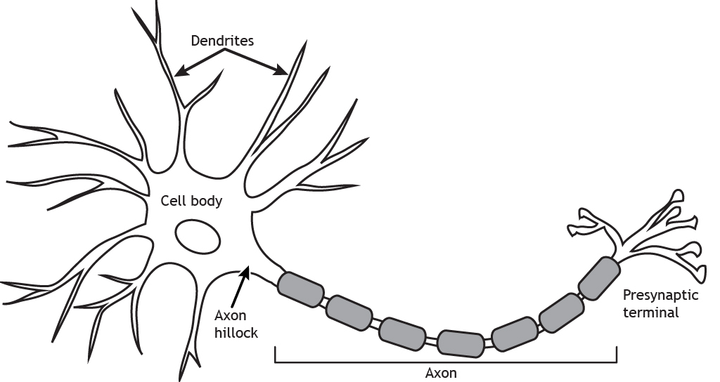
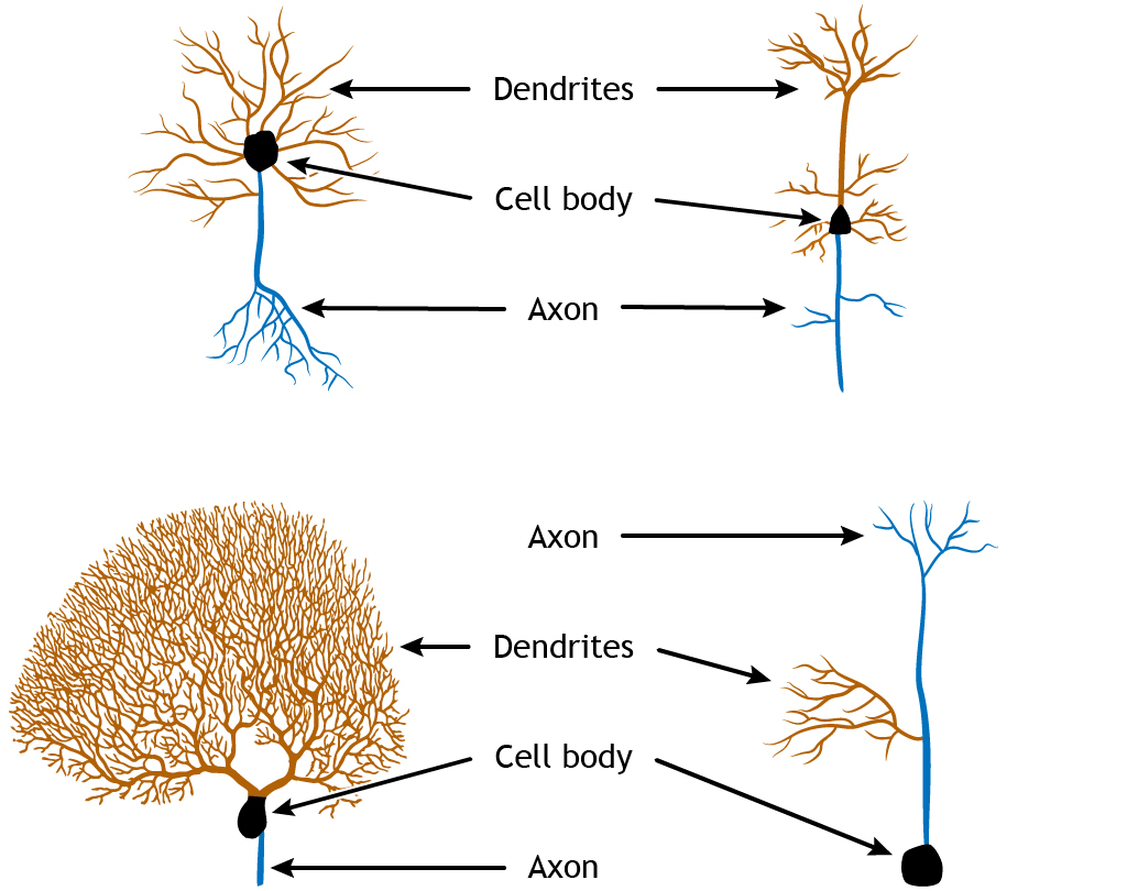
Neurons are the basic units of the brain. Their main function is to send electrical signals over short and long distances in the body, and they are electrically and chemically excitable. The function of the neuron is dependent on the structure of the neuron. The typical neuron consists of the dendrites, cell body, axon (including the axon hillock), and presynaptic terminal.
Although neurons do have a variety of adaptations that make them unique from other types of cells in the body, they are still cells. Therefore, they contain all of the basic features of a typical mammalian cell.
For example, they are made up of an aqueous cytoplasm bounded by a cell membrane. This cell membrane, also called a plasma membrane or lipid membrane, consists of a sheet of several individual molecules called phospholipids, which consist of two hydrophobic (water-fearing) tails and a hydrophilic (water-loving) end. These phospholipids arrange themselves into a bilayer, with the hydrophobic tails touching each other and the hydrophilic sides facing the cytoplasm and the extracellular space, which are both mostly water. Because of the chemical properties of the cell membrane, it is very effective at keeping ions and charged molecules separated, while allowing small molecules like water and oxygen across the cell.
Neurons also have all the organelles that you would see in other cell types, like a nucleus and mitochondria. The number of neurons in the adult human brain, according to our current best estimate, is close to 86 billion. This number was calculated using a revolutionary technique, the isotropic fractionator or “brain soup”, developed by Brazilian neuroanatomist Suzana Herculano-Houzel. To put this number in context, we have about 37 trillion cells in the whole body, so neurons in the brain make up about 0.2% of all cells in the body. Below are some unique characteristics that neurons have in common.
1. Neurons are electroactive, which means that they are charged cells that can change their charge.
2. Neurons are specialized for rapid communication.
Many cells are capable of sending and receiving chemical signals across long distances and time scales, but neurons are able to communicate with a combination of electrical and chemical signals in a matter of milliseconds. Additionally, the shape of neurons and the organization of the neurons on a microscopic level make them effective for sending signals in a very specific direction.
3. Neurons are “forever” cells.
We are constantly replacing non-neuronal cells. For example, the cells in our bones replace themselves frequently at a rate of about 10% each year. Our body makes new skin cells to replace the dying skin cells on the surface so that we have a “new” skin every month. The cells along the inside of our stomachs, exposed to very harsh acidic conditions, get replaced about every week. About 100 million new red blood cells are created every minute! On the other hand, the mature nervous system generally does not undergo much neurogenesis: the creation of new neurons.
The neurons that we have after development are the ones that we will keep until we die and this permanence of neuronal count makes them different from almost every other cell of the body. However, the idea of adult neurogenesis is a topic of debate among neuroscientists since some areas, like the olfactory system and the hippocampus, display new nerve cell production.
4. …But, neurons can change.
Even though new neurons are not created in most areas of the brain, neurons still have the capability to change in their structure and function. Some of these changes, such as physical changes to the structures of the input sites of the neurons, are believed to last for a lifetime.
We use the word plasticity to describe the ability for the brain to alter its morphology. This term is derived from the Greek plastikos, meaning “capable of being shaped or molded”—think of plastic surgery, where a person changes their physical appearance.
Also, neurons do have the capacity to repair themselves to some extent. Neurons of the Peripheral Nervous System may get injured or completely destroyed as a result of trauma to the body. Afterwards, those injured neurons can regrow to connect once again with their original partner. This regrowth seems to depend on a few chemical signals that the body produces, such as nerve growth factor and brain derived neurotrophic factor. However, this process is often very slow, and does not always successfully restore the nervous system to the way it was pre-injury.
Dendrites
The main function of neurons is to use changes in electrical properties in order to communicate with connected cells. This communication usually moves in one direction, and we will use this pathway as an outline for discussing the anatomical structures of the neurons.
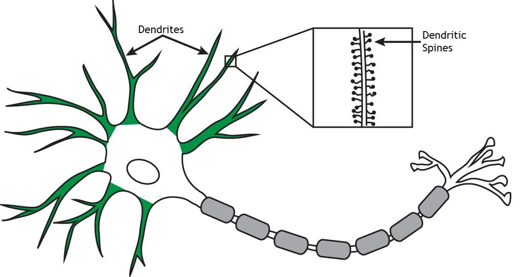
Dendrites, shown here in green, are processes that branch out in a tree-like fashion from the cell body. They are the main target for incoming signals received from other cells. The number of inputs a neuron receives depends on the complexity of the dendritic branching. Dendrites may also have small protrusions along the branches known as spines. Spines (illustrated in the inset box) are the sites of some synaptic contacts. Spines increase the surface area of the dendritic arbor, which may be an important factor in receiving communication.
We believe that spines are one of the most important sites where the nervous system is able to change. For example, neurons change shape after exposure to various environmental conditions, such as stress or exposure to drugs. Tiny changes to the surface of the neuron at the level of dendritic spines is an example of plasticity.
Dendritic plasticity is thought to underlie the reason that we can learn new facts or maintain memories about our childhood over long periods of time. Some set of tiny, submicroscopic changes to the morphology of dendritic spines may represent a single complex memory that you form. A neuron does not need spines for receiving information or for plasticity to take place. Many cells lack spines but are still capable of permanently changing. The input site may be anywhere along the dendrite, or even at the cell body—the “center” of the neuron.
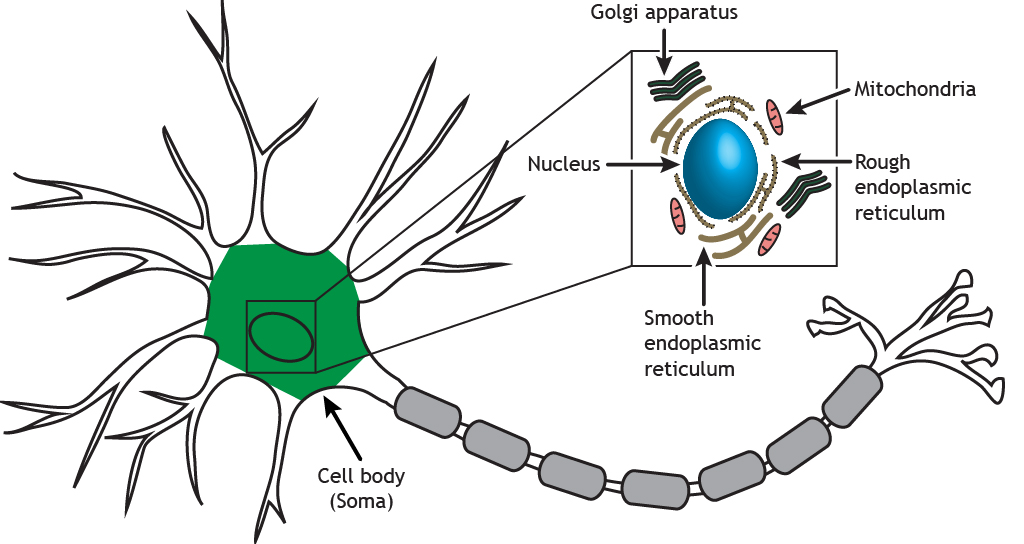
Cell Body
Information that arrives through the many dendrites of a neuron eventually filters into the cell body, or the soma, of the neuron. The cell body (shown below in green) contains the nucleus and cellular organelles, including the endoplasmic reticulum, Golgi apparatus, mitochondria, ribosomes, and secretory vesicles. The nucleus houses the DNA of the cell, which is the template for all proteins synthesized in the cell. The organelles (illustrated in the inset box) in the soma are responsible for cellular mechanisms like protein synthesis, packaging of molecules, and cellular respiration.
The cell body is responsible for deciding whether to pass a signal onto the next cell. The cell membrane of the soma performs a complex set of “cellular arithmetic” that weighs all of the incoming signals: excitatory, inhibitory, and modulatory signals. After all of the calculations have been performed, the membrane decides to send a signal, either a “yes” or “no” output, which travels down the axon.
Axon
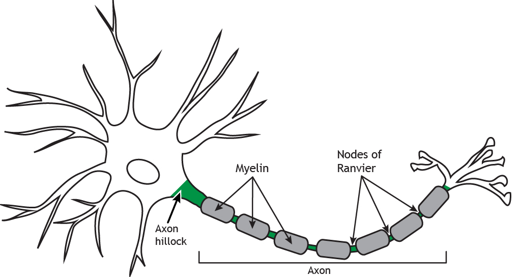
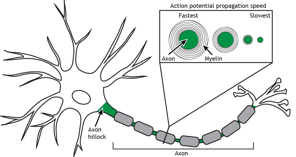
The axon is the main output extension of the neuron. The axon (highlighted in green) is usually a long, single process that begins at the axon hillock and extends out from the cell body. The axon hillock is located where the cell body transitions into the axon. Axons can branch in order to communicate with more than one target cell.
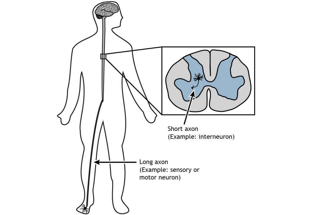
Several axons can bundle and travel together; these are nerves. Axons can be very long; the longest axon in the human body is part of the sciatic nerve that runs from the posterior end of the spinal cord down the leg to control the muscles of the big toe.

Action Potential
The axon transmits an electrical signal—called an action potential—from the axon hillock to the presynaptic terminal, where the electrical signal will result in a release of chemical neurotransmitters to communicate with the next cell. The action potential is a very brief change in the electrical potential, which is the difference in charge between the inside and outside of the cell. During the action potential, the electrical potential across the membrane moves from a negative value to a positive value and back.
Myelin
Many axons are also covered by a myelin sheath, a fatty substance that wraps around portions of the axon and increases action potential speed. There are breaks between the myelin segments called Nodes of Ranvier, and this uncovered region of the membrane regenerates the action potential as it propagates down the axon in a process called saltatory conduction. There is a high concentration of voltage-gated ion channels, which are necessary for the action potential to occur, in the Nodes of Ranvier.
Axon Characteristics
Axon Length
The length of an axon is variable depending on the location of the neuron and its function. The axon of a sensory neuron in your big toe needs to travel from your foot up to your spinal cord, whereas an interneuron in your spinal cord may only be a few hundred micrometers in length.
Axon Diameter
Axon diameter is also variable and can be used to differentiate different types of neurons. The diameter affects the speed at which the action potential will propagate. The larger the diameter, the faster the signal can travel. Additionally, larger diameter axons tend to have thicker myelin.
Axoplasmic Transport
Axoplasmic transport refers to the movement of material within the axon. Organelles, vesicles, and proteins can be moved from the cell body to the terminal via anterograde transport or from the terminal to the cell body via retrograde transport. Anterograde transport can be either fast or slow.
Microtubules run the length of the axon and provide the cytoskeleton tracks necessary for the transportation of materials. Proteins aid in axoplasmic transport. Kinesin is a motor protein that uses ATP and is used in anterograde transport of materials. Dynein is another motor protein that also uses ATP, but is used in retrograde transport of materials.
The Synapse
The synapse is the physical distance that separates two neurons.
Electrical Synapse
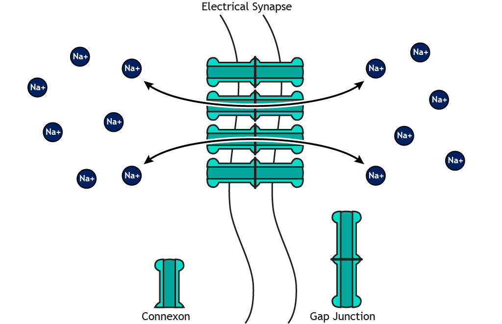
Electrical synapses physically share cytoplasm. An electrical synapse may be less than 5 nanometers apart. Cells connected by electrical synapses share cytoplasm, but have two separate cell membranes.
Chemical Synapse
Chemical synapses use neurotransmitters to communicate. Chemical synapses can vary depending on the nature of the synapse. A chemical synapse is a larger distance, about 15–40 nm across. Adjacent neurons connected by chemical synapses do not share cytoplasm.
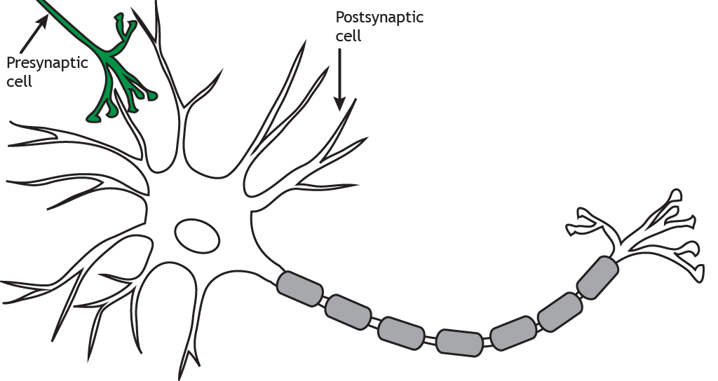
Presynaptic versus Postsynaptic
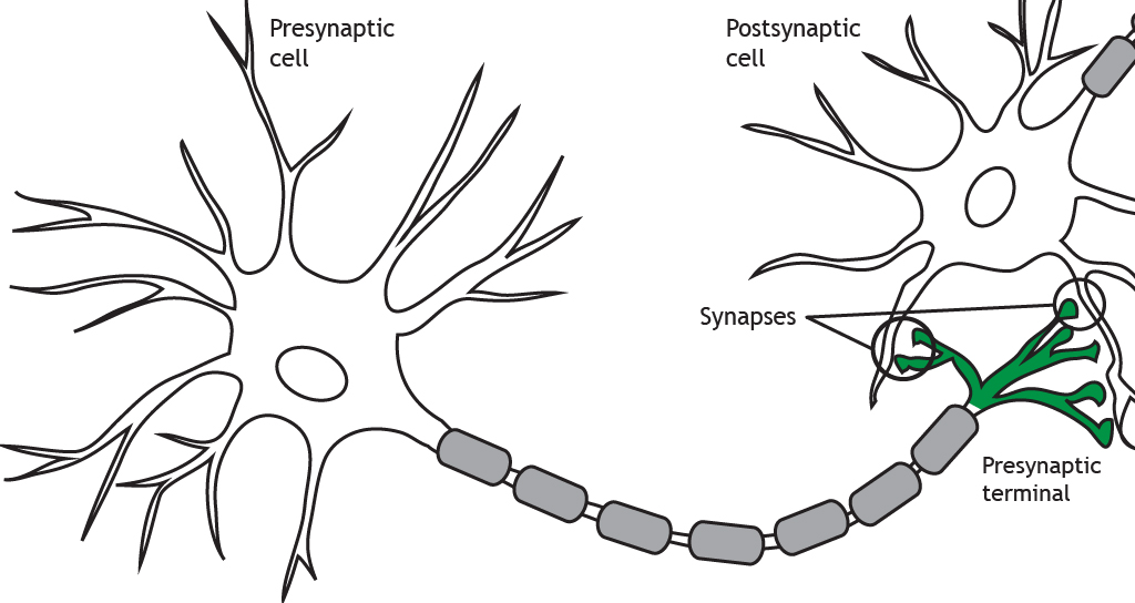
The axon terminates at the presynaptic terminal or terminal bouton. The terminal of the presynaptic cell forms a synapse with another neuron or cell, known as the postsynaptic cell. When the action potential reaches the presynaptic terminal, the neuron releases neurotransmitters into the synapse. The neurotransmitters act on the postsynaptic cell. Therefore, neuronal communication requires both an electrical signal (the action potential) and a chemical signal (the neurotransmitter). Most commonly, presynaptic terminals contact dendrites, but terminals can also communicate with cell bodies or even axons. Neurons can also synapse on non-neuronal cells such as muscle cells or glands.
The terms presynaptic and postsynaptic are in reference to which neuron is releasing neurotransmitters and which is receiving them. Presynaptic cells release neurotransmitters into the synapse and those neurotransmitters act on the postsynaptic cell.
Variations in Structure
Although these typical structural components can be seen in all neurons, the overall structure can vary drastically depending on the location and function of the neuron. Some neurons, called unipolar, have only one branch from the cell body, and the dendrites and axon terminals project from it. Others, called bipolar, have one axonal branch and one dendritic branch. Multipolar neurons can have many processes branching from the cell body. Additionally, each of the projections can take many forms, with different branching characteristics. The common features of cell body, dendrites, and axon, though, are common among all neurons.
Key Takeaways
- Each structural component of the neuron has an important function
- Overall structure of the cell can vary depending on location and function of the neuron
Test Yourself!
Attributions
Portions of this chapter were remixed and revised from the following sources:
- Foundations of Neuroscience by Casey Henley. The original work is licensed under a Creative Commons Attribution-NonCommercial-ShareAlike 4.0 International License
- Open Neuroscience Initiative by Austin Lim. The original work is licensed under a Creative Commons Attribution-NonCommercial 4.0 International License.
Electrically excitable cells of the nervous system that receive and transmit signals to different areas of the body.
Non-neuronal cells that are not electrically excitable.
Processes that branch out in a tree-like fashion from the neuronal cell body and are the main target for incoming signals received from other neurons.
Small protrusions off dendrites that serve as synaptic site
The ability to change
The cell body of a neuron. Where the nucleus and organelles are located.
The main extension off the cell body of a neuron that provides output of the cell.
A bundle of axons in the peripheral nervous system
The electrical signal transmitted by the axon of a neuron.
The junction of the cell body and the axon. Where action potentials are generated.
Chemicals that are released by neurons or other cells that bind to receptors on other neurons or cells to elicit change in the target cell.
The electrical charge of the neuron (measured in mV).
fatty substance that covers the axons of some neurons
Breaks between adjacent sheaths of myelin on the neuron axon.
a straight line passing from side to side through the center of a body or figure, especially a circle or sphere.
Movement from the cell body to the axon terminals
Movement from the axon terminals toward the cell body
A motor protein that uses ATP in anterograde transport of materials
Motor protein that uses ATP in retrograde transport of materials
Junction of two neurons or a neuron and a target cell.
Synapse where the cytoplasm of 2 cells is physically shared.
A synapse where 2 cells are separated by a synaptic cleft that chemical neurotransmitters must cross to signal between cells.
The end of the axon furthest from the cell body.
Release neurotransmitters into the synapse
Cells that receive neurotransmitters
