
5 Ion Movement
Ion flow into and out of the neuron is a critical component of neuron function. Ions move in predictable ways, and the control of ion movement affects the cell at rest and while sending and receiving information from other neurons.
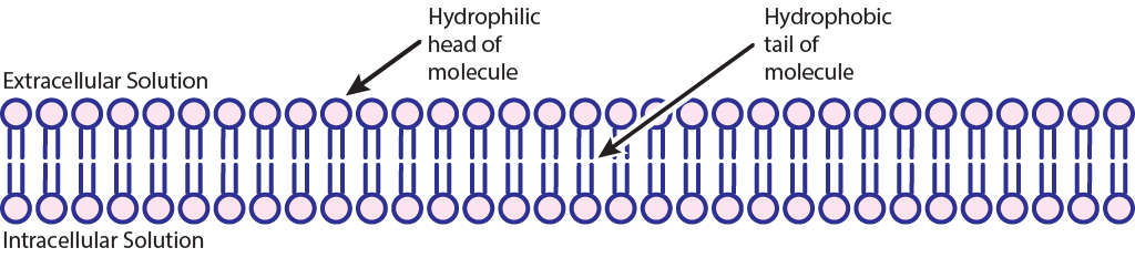
Phospholipid Bilayer Prevents Ion Movement
The cell membrane that separates the inside of the cell from the outside is a very effective boundary. It is described as being selectively permeable, which means that some molecules are able to travel across the membrane easily, other molecules have an intermediate ability to cross, and other molecules are completely incapable of passing. Generally, gases and molecules of water are able to pass through the cell membrane easily. Large molecules like glucose, and charged molecules like ions or amino acids, are unable to pass across the membrane.
The neuronal membrane is composed of lipid molecules that form two layers. The hydrophilic heads of the molecules align on the outside of the membrane, interacting with the intra- and extracellular solution of the cell, whereas the hydrophobic tails are arranged in the middle, forming a barrier to water and water-soluble molecules like ions. This barrier is critical to neuron function.
Ion Channels Allow Ion Movement
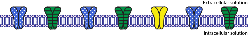
Most cells of the body, including neurons, have specialized transmembrane proteins embedded in the cell membrane. These transmembrane proteins are huge protein complexes that span the entirety of the membrane, with an outer side and an inner side. In the middle of the protein is a pore, which is essentially a “tunnel” that allows molecules and ions to pass across the cell membrane. These proteins are called ion channels. These channels are passive since they do not use any cellular energy to move ions. Rather, they simply provide easy passage for ions. It may be useful to think of an ion channel as a “cellular door”. Ion channels are embedded throughout the neuronal membrane.
Channels can be opened and closed in a number of different ways. We can categorize ion channels into four major classes based on their opening and closing conditions.
- Leak channels are persistently open. You can think of these leak channels as revolving doors that are never locked. Neurons usually have several leak channels.
- Voltage-gated ion channels open in response to a change in membrane potential.
- Ligand-gated ion channels open in response to chemical (ligand) binding, such as neurotransmitters.
- The fourth class of ion channels is a catch-all category that includes a wide variety of channels that are used by the sensory systems. They open and close in response to unique stimuli depending on what they are able to sense. For example, some open and close depending when they are moved physically, such as a distortion or stretch (mechanoreceptors). We have these in the hair cells of our ears, in our skin, and in our muscles. Photoreceptors in our eyes have ion channels that close in response to being hit by photons of light, and this activity is necessary for us to be able to see in both brightly and dimly lit environments.
One important feature of ion channels is their ability to distinguish ions based on their chemical properties. For example, some channels are selective for Na+, while preventing the passage of all other ions. Each ion channel has special molecular characteristics that allow certain types of ions to pass through the pore while excluding other ions. Channels can be specific to one ion or allow the flow of multiple ions.
Ion channels control ion movement across the cell membrane because the phospholipid bilayer is impermeable to the charged atoms. When the channels are closed, no ions can move into or out of the cell. When ion channels open, however, then ions can move across the cell membrane.
Gradients Drive Ion Movement
Ions move in predictable ways. Concentration (chemical) and electrical gradients drive ion movement. The chemical gradient refers to the natural process by which a high concentration of a substance, given enough time, will eventually diffuse to a lower concentration and settle evenly over the space. Ions will diffuse from regions of high concentration to regions of low concentration. Diffusion is a passive process, meaning it does not require energy. As long as a pathway exists (like through open ion channels), the ions will move down the concentration gradient.
In addition to concentration gradients, electrical gradients can also drive ion movement. The electrical gradient refers to the electrical forces acting on charged molecules, “pulling” opposite charges together while also “pushing” like charges away from one another—just like the polarity of magnets. Ions are attracted to, and will move toward, regions of opposite charge. Positive ions will move toward regions of negative charge, and vice versa.
For discussion of ion movement in this text, the combination of these two gradients will be referred to as the electrochemical gradient. Sometimes the concentration and electrical gradients driving ion movement can be in the same direction; sometimes the direction is opposite. The electrochemical gradient is the summation of the two individual gradients and provides a single direction for ion movement.
When Gradients Balance, Equilibrium Occurs
When the concentration and electrical gradients for a given ion balance—meaning they are equal in strength, but in different directions—that ion will be at equilibrium. Ions still move across the membrane through open channels when at equilibrium, but there is no net movement in either direction, meaning there is an equal number of ions moving into the cell as there are moving out of the cell.
Important Ions for Neurons
Sodium, potassium, and chloride ions are found in different concentrations across the neuron cell membrane. The location of these ions across the cell membrane and their concentration gradients are important for the function of the neuron. Sodium (Na+) ions are more concentrated in the extracellular fluid and less concentrated within the intracellular fluid. Whereas potassium (K+) ions are more concentrated within the intracellular fluid and less concentrated in the extracellular fluid.
| Ion | Inside concentration (mM) | Outside concentration (mM) |
|---|---|---|
| Sodium (Na+) | 15 | 145 |
| Potassium (K+) | 125 | 5 |
| Chloride (Cl-) | 13 | 150 |
Key Takeaways
- The phospholipid bilayer prevents ion movement into or out of the cell
- Ion channels allow ion movement across the membrane
- Electrochemical gradients drive the direction of ion flow
- At equilibrium, there is no net ion movement (but ions are still moving)
Test Yourself!
Attributions
Portions of this chapter were remixed and revised from the following sources:
- Foundations of Neuroscience by Casey Henley. The original work is licensed under a Creative Commons Attribution-NonCommercial-ShareAlike 4.0 International License
- Open Neuroscience Initiative by Austin Lim. The original work is licensed under a Creative Commons Attribution-NonCommercial 4.0 International License.
Transmembrane proteins that have a pore that allow for the passage of ions between the inside and outside of the cell
Ion channels that are always open
Ion channels that open/close in response to changes in voltage (membrane charge)
Ion channels that open in response to the binding of a chemical ligand (such as a neurotransmitter)
Ion channels that open in response to physical distortion
Channels that open in response to light
Does not allow passage
Process by which a high concentration of a substance, given enough time, will eventually diffuse to a lower concentration and settle evenly over the space
Passive process when substances move from a higher concentration to a lower concentration
electrical forces acting on charged molecules, “pulling” opposite charges together while also “pushing” like charges away from one another
The combination of chemical and electrical gradients
