
67 Molecular Mechanisms of Memory: Aplysia
Through the 1960s, psychiatrist Eric Kandel and his colleagues Arvid Carlsson and Paul Greengard worked with the marine mollusk Aplysia californica. Their work uncovering the neural mechanisms of learning and memory earned them the Nobel Prize in Physiology or Medicine in 2000.
With a nervous system of only 20,000 cells, Aplysia is orders of magnitude simpler than the other model organisms used at the time. Additionally, some Aplysia neurons are huge, up to a millimeter in diameter, which took away the need for highly precise equipment.

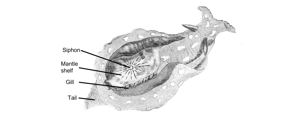
Aplysia also has a relatively simple anatomy. It breathes using a half-circle of delicate tissue called the gill, which is guarded by the mantle shelf. They also have an organ called the siphon, a small tube that is used for moving water through the animal.
Kandel and his colleagues began their exploration of memory by studying the gill-withdrawal reflex, a defensive motor response behavior. When a stimulus, such as a hungry predator (or an experimenter’s paintbrush), grazed the siphon, the Aplysia would reflexively withdraw their gill, as if to protect this vital organ by shrinking away from the threat. However, after repeated brush strokes to the siphon, the sea slugs figured out that the stimulus was completely innocuous and decreased the strength of gill withdrawal. Kandel and team suggested that this change in behavior was a form of learning. Kandel and colleagues used Aplysia to learn about the processes of habituation and sensitization.
Habituation
The gill-withdrawal reflex circuit relies heavily on two different populations of neurons: the glutamatergic sensory neurons that receive somatosensory information from the skin of the siphon, and the motor neurons that control the muscles of the gill. By using two different tiny glass pipettes, they could impale these neurons, inducing action potential firing in the sensory neuron, and observe changes in membrane potential of the motor neurons. Activation of the sensory neuron causes release of glutamate onto the motor neuron that controls the gill, binding to glutamatergic receptors and causing an EPSP response in the motor neuron.
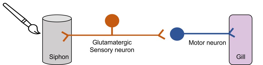
Although there was a strong gill withdrawal reflex following the one poke, Kandel and colleagues observed that after repeated pokes to the siphon, the gill withdrawal reflex got weaker. They found that the activity of the siphon neuron remained unchanged, however, the activity of the motor neuron supplying the gill decreased. The reflex had been habituated. This habituation was due to a decreased release of glutamate from the siphon sensory neuron onto the gill motor neuron.
Sensitization
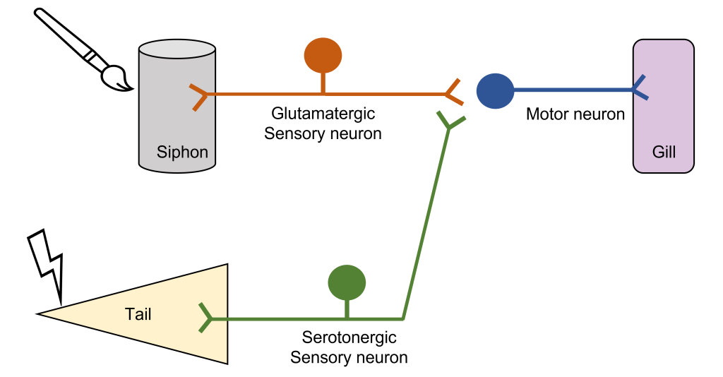
The group went about seeing if they could modify this habituated response, curious if a stored memory can be modified by stimuli from the outside world. When they paired the mild siphon touch with a painful electric shock to the tail, the Aplysia began responding with a strong motor reaction, withdrawing the gill very intensely, indicating that the inhibited response disappeared. They called this observation sensitization. In electrophysiological studies, they observed that the EPSP at the motor neuron was much larger following the tail shock.
On a molecular level, presentation of sensitization is downstream of the action of a third population of neurons, serotonergic interneurons that synapse onto the siphon sensory neurons. The noxious stimulus triggers these interneurons to release the neurotransmitter serotonin, which binds to receptors on the terminals of the siphon sensory neurons, increasing adenylyl cyclase activity and cAMP production. cAMP activates protein kinase A, which in turn increases the release of glutamate from the sensory neuron and strengthening the gill withdrawal reflex.
Repeating the sensitization protocol multiple times results in plastic changes at the synapse between the sensory siphon neuron and the motor neuron. These plastic changes are long-lasting and dependent on new gene expression. The activated protein kinase A can translocate to the siphon sensory neuron nucleus to activate gene expression and the production of proteins that can remodel the anatomy of the synapse through the growth of additional sensory neuron axons, increasing synaptic connections.
Key Takeaways
- Aplysia is a simple animal model that has been used to study learning and memory.
- Touching the siphon activates the gill withdrawal reflex through the release of glutamate on to the gill motor neuron.
- Repeatedly touching the siphon results in habituation of the gill withdrawal reflex, and a weaker gill withdrawal.
- Shocking the tail following habituation activates a serotonergic neuron that facilitates glutamate release from from the siphon neuron, producing a strong gill withdrawal reflex.
- Over the long-term, this produces long lasting plasticity within this synapse.
Attributions
Portions of this chapter were remixed and revised from the following sources:
- Open Neuroscience Initiative by Austin Lim. The original work is licensed under a Creative Commons Attribution-NonCommercial 4.0 International License.
Media Attributions
- aplysia © Columbia University, New York adapted by Valerie Hedges is licensed under a Public Domain license
- aplysia anatomy © Phillip Henry Gosse adapted by Valerie Hedges is licensed under a Public Domain license
- Synapse between the siphon and the gill © Valerie Hedges is licensed under a CC BY-NC-SA (Attribution NonCommercial ShareAlike) license
- Synapse between siphon, tail and gill © Valerie Hedges is licensed under a CC BY-NC-SA (Attribution NonCommercial ShareAlike) license
decrease in response after repeated presentation of a stimulus
increased effect of a drug after repeated administration
