
27 Peripheral Nervous System
The Peripheral Nervous System (PNS) functions as the intermediary between the central nervous system (CNS) and the rest of the body, including the skin, internal organs, and muscles of our limbs. The PNS can be divided into three main branches:
- Somatic nervous system
- Autonomic nervous system
- Enteric nervous system
In this chapter, the somatic and autonomic nervous systems will be compared and contrasted. An overview of the enteric nervous system will also be provided.
Somatic vs. Autonomic Nervous System
The somatic and autonomic nervous system differ in their:
- Target/effector organs
- Efferent pathways and the neurotransmitters that are used
- How the target/effector organ responds to the neurotransmitter
Peripheral Nervous System: Somatic Nervous System
The somatic nervous system represents all the parts of the PNS that are involved with the outside environment, either in sensing the environment or acting on it. For example, the nerves that detect pressure or pain on the foot are part of the afferent somatic nervous system. We also think of the somatic nervous system as the branch that sends signals to our skeletal muscles. The nerves that innervate the muscles of the legs as we run are part of the efferent somatic nervous system. The somatic nervous system is also called the “voluntary nervous system” since it is used to cause muscle movement related to intentional actions.
The somatic nervous system has an efferent path from the central nervous system to the target / effector organ that is made up of one neuron. The axon of this neuron is heavily myelinated, which allows for fast delivery of messages. This somatic motor neuron has its cell body in the central nervous system and has an axon that extends all the way to the target/effector organ, which—for the somatic nervous system—will be a skeletal muscle.
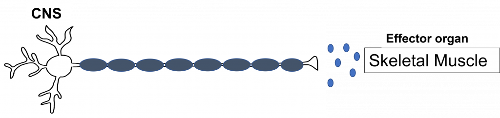
Synapse: Neuromuscular Junction
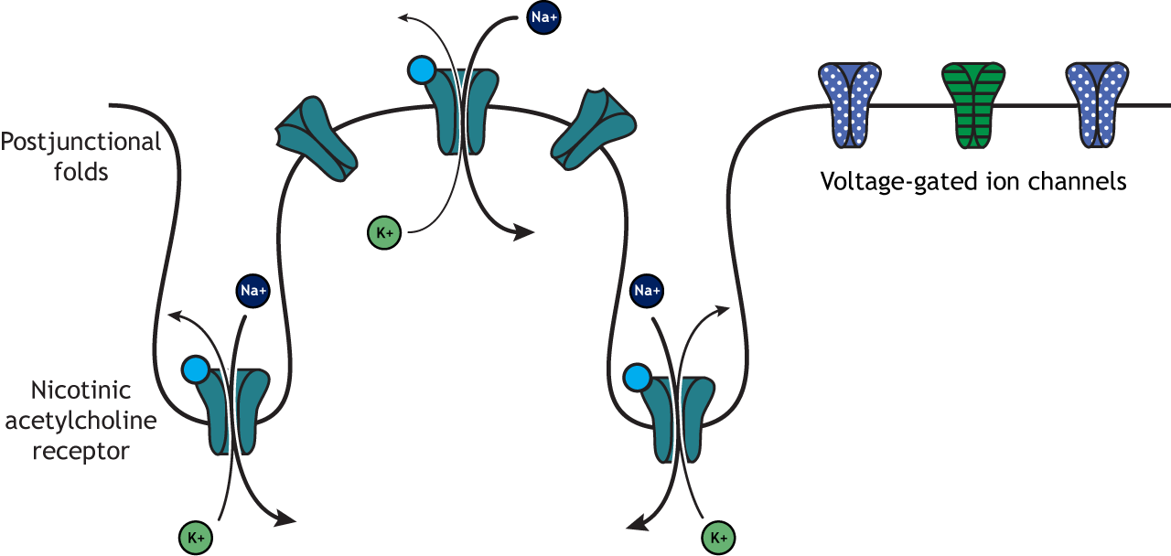
All somatic motor neurons release the same neurotransmitter, acetylcholine, onto the skeletal muscle target/effector organ at the neuromuscular junction (NMJ). The NMJ is one of the largest synapses in the body and one of the most well-studied because of its peripheral location. Acetylcholine is the neurotransmitter released at the NMJ and it acts upon ligand-gated, non-selective cation channels called nicotinic acetylcholine receptors that are present in postjunctional folds of the muscle fiber. Acetylcholinesterase, an enzyme that breaks down acetylcholine and terminates its action, is present in the synaptic cleft of the neuromuscular junction.
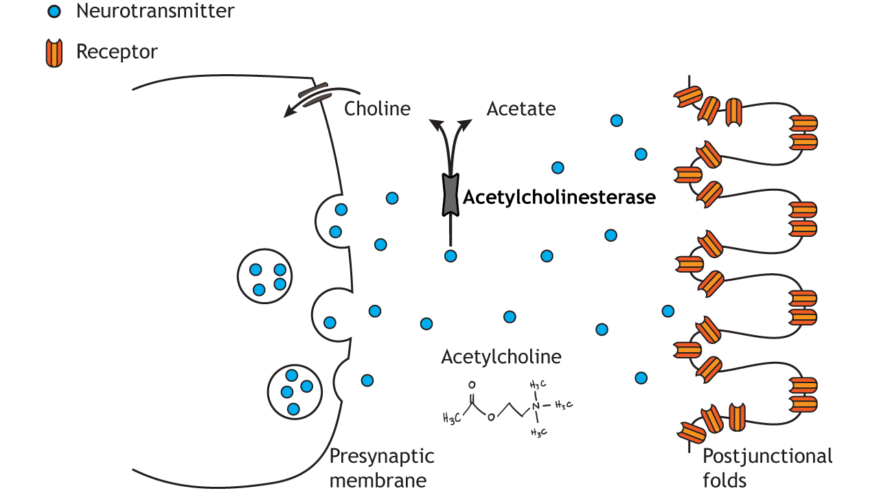
Nicotinic acetylcholine receptors allow for the influx of sodium ions into the muscle cell. The depolarization will cause nearby voltage-gated channels to open and fire an action potential in the muscle fiber. In a healthy system, an action potential in the motor neurons always causes an action potential in the muscle cell. The action potential leads to contraction of the muscle fiber.
Peripheral Nervous System: Autonomic Nervous System
The autonomic nervous system encompasses all the branches of the peripheral nervous system that deal with the internal environment. As with the somatic nervous system, the autonomic nervous system is comprised of nerves that detect the internal state as well as nerves that influence the internal organs. The body carries out all sorts of functions and responses unconsciously without any intentional control. It can do so by sending signals to smooth muscles and glands. The signals that cause us to sweat when it is hot, our pupils to dilate when it is dark, and our blood pressure to adjust when we stand up too quickly are all driven by the nerves of the autonomic nervous system.
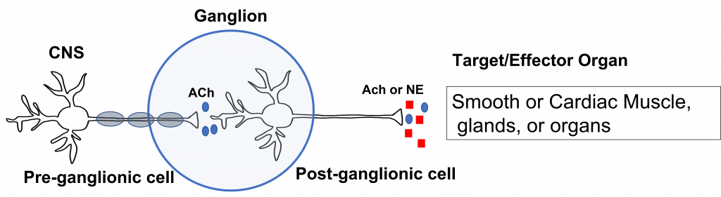
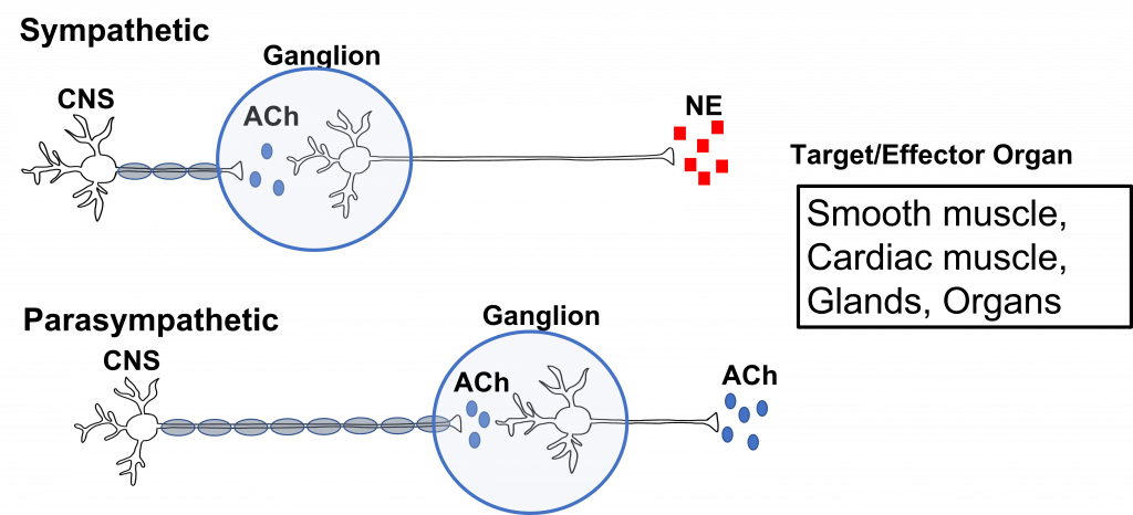
The autonomic nervous system has an efferent path from the central nervous system to the target/effector organ that is made up of a chain of two neurons with a ganglion in the middle. Recall that a ganglion is a collection of neuron cell bodies in the periphery.
The neuron that has its cell body in the central nervous system is called the preganglionic neuron. The preganglionic cell axon is lightly myelinated and extends to a ganglion in the periphery where it synapses on the second neuron in the chain called the postganglionic neuron. The postganglionic neuron has its cell body in the ganglion and its unmyelinated axon extends from the ganglion to the target/effector organ, which for the autonomic nervous system will be: smooth muscle, cardiac muscle, glands, or organs.
The first synapse happens at the site of the ganglion. All preganglionic cells release acetylcholine onto the postganglionic cells. The acetylcholine binds to nicotinic acetylcholine receptors, which results in the generation of an excitatory postsynaptic potential for the postganglionic cell body. This effectively passes the message between the preganglionic and postganglionic cells.
The second synapse occurs between the postganglionic cell and the target/effector organ. Postganglionic neurons in the autonomic nervous system release either norepinephrine or acetylcholine (more on this later). These neurotransmitters can have either stimulatory or inhibitory activity at the target organs, dependent on the properties of the postsynaptic receptor that they bind to.
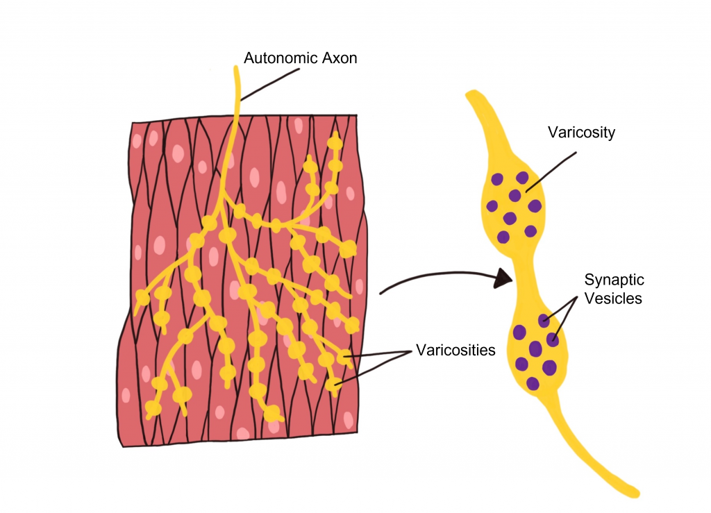
Autonomic Effector Synapse
Within the autonomic nervous system, synapses have a different structure than what is observed at the NMJ of the somatic nervous system. Instead, autonomic axons are highly branched and have enlarged varicosities that are spread along the axon. These varicosities contain synaptic vesicles that are filled with neurotransmitters. The varicosities form synapses ‘en passant’ (literally meaning ‘in passage’) with the target organ. These axon branches with varicosities drape over the cells of the target tissue, allowing a single axon branch to affect change over a greater area of the target tissue and better distribute autonomic activation at the target tissue.
Comparing Somatic to Autonomic Nervous System
In summary, the somatic nervous system has a one neuron path that originates in the central nervous system. That neuron extends all the way to the skeletal muscle (target tissue) where it releases acetylcholine. The acetylcholine binds to nicotinic acetylcholine receptors that cause EPSPs in the muscle fibers and giving a stimulatory effect. The autonomic nervous system (both branches) use a two neuron pathway between the CNS and the target tissues that are smooth muscle, cardiac muscle, glands, and organs. The sympathetic division consists of short presynaptic neurons that synapse on longer postsynaptic neurons in peripheral ganglia that are situated close to the spinal cord. The parasympathetic division consists of long presynaptic neurons that synapse on shorter postsynaptic neurons in peripheral ganglia that are situated close to the target organ. Like the somatic motor system, the primary neurotransmitter in the parasympathetic division is acetylcholine. In the sympathetic division, the preganglionic neuron releases acetylcholine, but the postganglionic neuron releases norepinephrine. The effects of these neurotransmitters at the target tissue can be either stimulatory or inhibitory depending on the properties of the postsynaptic receptor. This information is summarized in the figure and table below.
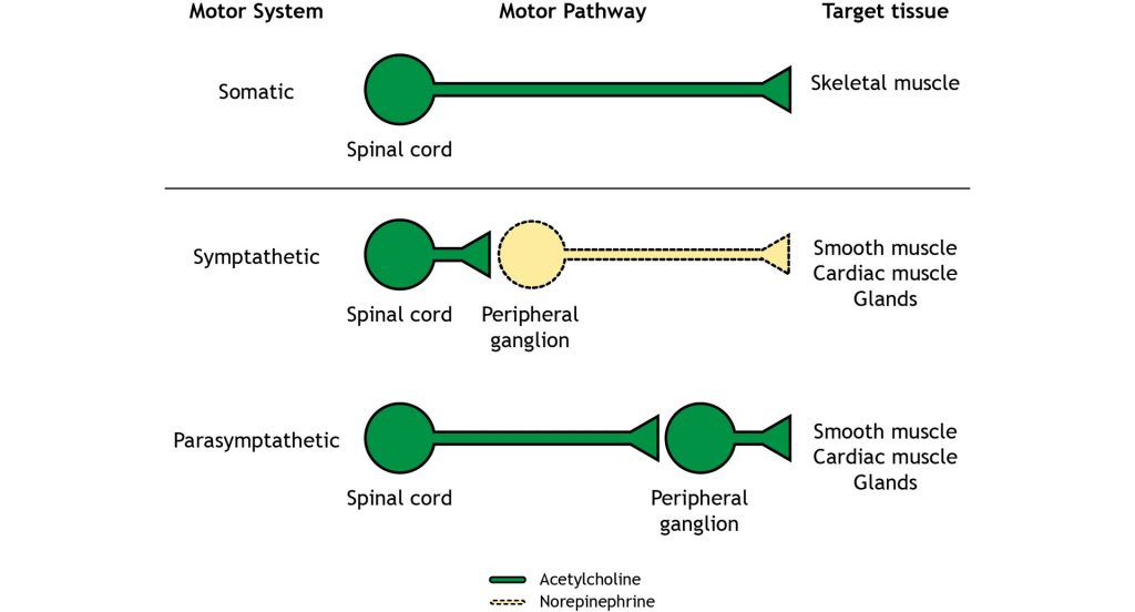
| Branch of PNS | Efferent Path | Target / Effector Organ | Neurotransmitter Released at Ganglion and Effect | Neurotransmitter Released at Target Organ | Target Organ Response to Neurotransmitter |
|---|---|---|---|---|---|
| Somatic | 1 Neuron | Skeletal Muscle | None- No Ganglion | Acetylcholine | Stimulatory |
| Autonomic | 2 Neuron Chain | Smooth / Cardiac Muscle, Glands, Organs | Acetylcholine, excitatory | Acetylcholine or Norepinephrine | Can be stimulatory or inhibitory |
Two Branches of the Autonomic Nervous System
There are two branches of the autonomic nervous system, called the sympathetic nervous system and the parasympathetic nervous system, that typically have opposite effects at target tissues.
Consider a scenario in which you encounter a bear on hike through Yellowstone National Park. Your heartrate would likely increase and your breathing rate would increase as you contemplate how to survive your encounter. This complex set of physiological reactions are due to activation of one of the branches of the autonomic nervous system: the sympathetic nervous system. The sympathetic nervous system mobilizes a set of physiological changes to a threat that is sometimes called the fight-or-flight response, which is activated when we are faced with a threat, either perceived or real. All of these rapid bodily responses result in the body preparing to attack or defend itself. Increased respiration allows the body to take in more oxygen, and dilation of blood vessels in the muscles allows that oxygen to get to the muscles, which is needed for muscle activation.
Now, consider a completely opposite scenario. You’ve just eaten a large meal and you are spending the evening watching television on your couch. You would probably feel relaxed, satisfied, and more than a little sluggish. A different physiological response is happening, a behavior called the rest-and-digest response. These physiological changes are driven by the other main branch of the autonomic nervous system, called the parasympathetic nervous system.
Targets and Effects of the 2 Branches of the Autonomic Nervous System
The sympathetic nervous system and the parasympathetic nervous system are referred to as antagonistic. These branches are called ‘antagonistic’ because they typically have opposite effects at target tissues.
There is dual innervation at many target organs of the autonomic nervous system. This means that target organs receive connections from both sympathetic and parasympathetic neurons. In a sense, this dual innervation provides both a ‘break’ and an ‘accelerator’ for changing the activity of our internal organs, offering a good amount of control.
Both the sympathetic nervous system and parasympathetic nervous systems influence the internal organs simultaneously. At all times, the heart is getting signals from the sympathetic nervous system which increase heart rate, and signals from the parasympathetic nervous system which decreases heart rate. However, this seesaw-like balance can shift quickly in either direction, such as inducing a sympathetic response if a fearful stimulus is encountered.
| Target Organ | Sympathetic Action | Parasympathetic Action |
|---|---|---|
| Eye | Pupil dilation | Pupil constriction |
| Salivary glands | Prevents saliva secretion | Increases saliva secretion |
| Lungs | Airway dilation | Airway constriction |
| Heart | Increases heart rate | Decreases heart rate |
| Blood vessels | Constriction | No innervation |
| Stomach, liver, intestines, pancreas | Decreases digestion | Increases digestion |
| Kidneys | Sodium and water retention | No innervation |
| Adrenal gland | Increases epinephrine and norepinephrine secretion | No innervation |
| Reproductive organs | Orgasm and ejaculation | Blood vessel dilation leading to erection |
Anatomical Differences between 2 Branches of Autonomic Nervous System
The sympathetic and parasympathetic nervous systems differ anatomically as well. The sympathetic preganglionic neurons are short and the sympathetic postganglionic neurons are long. The parasympathetic preganglionic neurons are long and the parasympathetic postganglionic neurons are short.
Sympathetic Anatomy
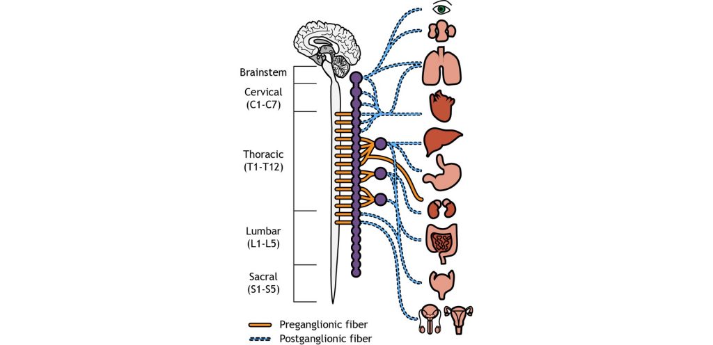
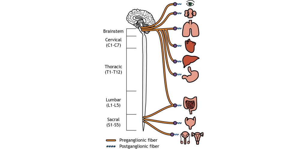
The cell bodies of the preganglionic neurons of the sympathetic nervous system are located in the thoracic and lumbar sections of the spinal cord, so we can use thoracolumbar to describe the site of origin. The axons leave the central nervous system and travel only a short distance to the peripheral ganglion. In the sympathetic system, most of the ganglia are located along the spinal cord in the sympathetic paravertebral chain. A few sympathetic ganglia, called prevertebral, are located slightly more laterally. The preganglionic neurons synapse on the postganglionic neurons in the sympathetic ganglia. The postganglionic neurons then travel the rest of the distance to synapse on the target organs.
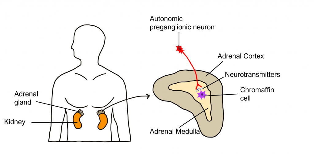
One exception to this structure is the innervation of the adrenal medulla. The adrenal gland is a structure located at the top of each kidney. The gland has an outer portion called the cortex and an inner portion called the adrenal medulla, which is made up of cells called chromaffin cells. The sympathetic preganglionic cell releases acetylcholine at the synapse with the chromaffin cells of the medulla and releases acetylcholine onto the chromaffin cell that is serving as a modified post-ganglionic neuron. Then the chromaffin cells of the adrenal medulla release epinephrine and norepinephrine into the bloodstream. Within the bloodstream these neurotransmitters can act as hormones affecting change throughout the body leading to a rapid onset of sympathetic activity and sustained activation.
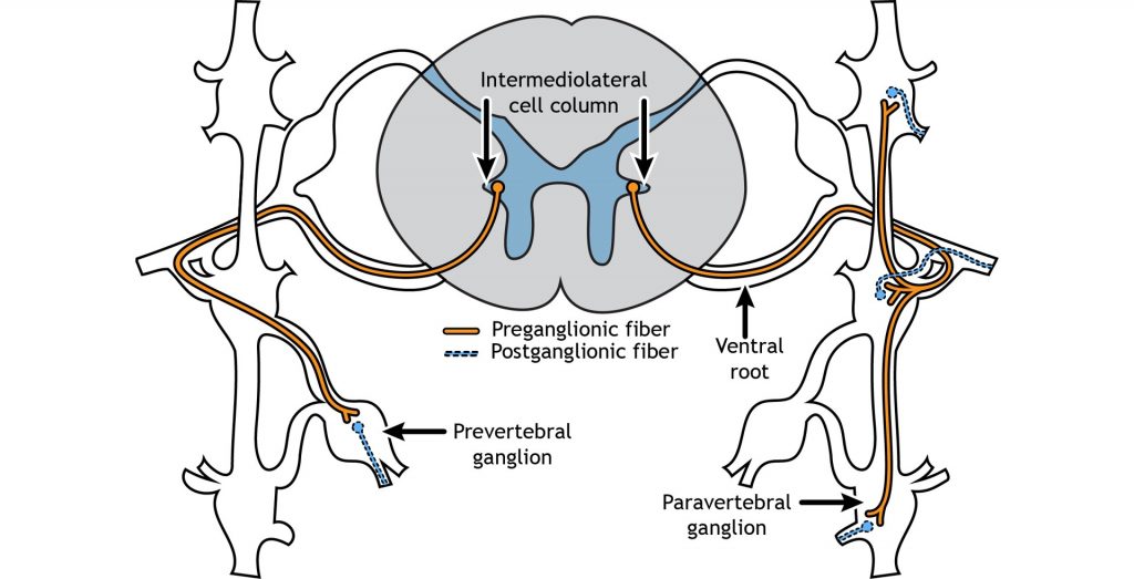
The sympathetic preganglionic cell bodies are located in the intermediolateral cell columns in the lateral horn of the spinal cord, a gray matter region between the dorsal and ventral horns. The neurons send their axons out through the ventral root, and then can take one of multiple pathways. The axon can terminate in the paravertebral ganglion at the same spinal level as the cell body, or the axon can extend up or down the sympathetic chain to synapse on a postganglionic fiber at a different spinal level. Another possible pathway is to enter and exit the sympathetic chain without synapsing and continue on to terminate in a prevertebral ganglion located closer to the target organ.
Parasympathetic Anatomy
On the other hand, parasympathetic neurons originates predominantly in the cervical spinal cord (near the neck), with some signals originating in the sacral areas (near the tail bone). The parasympathetic nervous system usually receives signals from several cranial nerves. Cranial nerve X, also called the vagus nerve innervates multiple bodily organs in the midsection of the body. For the parasympathetic nervous system, the ganglia are located very close to the target tissues. Therefore, the preganglionic neuron must be very long to reach all the way to the ganglion to makes its first synapse. But due to the proximity of the ganglion to the target tissue, the postganglionic neuron is very short.
Neurotransmitters of the Autonomic Nervous System
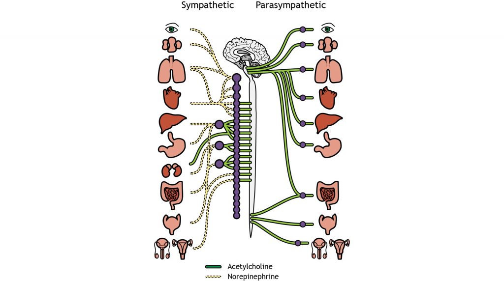
We have already established that at the first synapse in the chain of autonomic neurons, all preganglionic neurons release acetylcholine, which binds to excitatory nicotinic receptors on the postganglionic neurons. This is true of both the sympathetic and parasympathetic nervous system. However, the two branches of the autonomic nervous system have different effects at target tissues because they use different neurotransmitters that produce either stimulatory or inhibitory effects at target tissues.
The sympathetic postganglionic neurons release norepinephrine at target tissues. Norepinephrine (and epinephrine) binds two major classes of adrenergic receptors: alpha adrenergic receptors and beta adrenergic receptors. The effects of norepinephrine (and epinephrine) can be either excitatory or inhibitory depending on the properties of the postsynaptic receptor.
The parasympathetic postganglionic neurons release acetylcholine at target tissues. The target tissues express muscarinic acetylcholine receptors. The effect of the acetylcholine can be either excitatory or inhibitory depending on the subtype of muscarinic (metabotropic) receptors. The location of function of these transmitters and receptors are critical for clinical use, for example, when treating high blood pressure or sexual dysfunction.
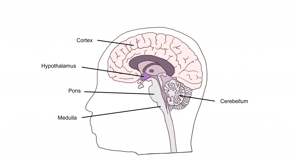
Autonomic Control Centers in the Brain
Visceral functions are regulated through a number of autonomic reflexes, but they can be directly controlled by higher brain areas. Within the brain stem, the medulla most directly controls autonomic activity. The medulla receives most of its information via the vagus nerve (a mixed nerve that contains both sensory and motor fibers). Within the medulla are a variety of control centers that regulate functions such as cardiovascular activity, respiratory rate, vomiting, and swallowing. The pons, another brain stem structure also serves as an autonomic control center for respiration.
In addition to brain stem structures, there are also higher order brain areas that are important for autonomic control. The hypothalamus can directly regulate the medulla, and is also critical for the regulation of water balance, temperature control, and hunger. The Limbic System, made up of a number of structures including the the hippocampus and amygdala, functions in our expression of emotion. The limbic system structures are responsible for visceral responses associated with emotional states, including blushing, fainting, pallor, nervous cold sweats, racing heart rate, and uneasy feelings in the stomach.
Further, the cerebral cortex and cerebellum also function in autonomic control. The cerebral cortex has been shown to regulate lower brain structures especially in autonomic control associated with emotion and personality. The cerebellum has connections to the medulla that are critical for the control of autonomic functions such as sweating and nausea.
Enteric Nervous System
The internal organs that carry out digestive functions, such as the esophagus, stomach, and intestines, are surrounded by a dense mesh of neurons that regulate their activity. Consisting of half a billion nerve cells, this net of neurons cause the digestive tract to increase or decrease the rate of these processes depending on the body’s demands The enteric nervous system receives signals from both the sympathetic and parasympathetic nervous systems, and functions without our conscious knowledge. Historically, these digestive functions have been classified as part of the autonomic nervous system, but these responses do not share the same reflex pathway, and the enteric signals can work entirely independent of the vagus nerve, for example.
Key Takeaways
- There are 3 main divisions of the peripheral nervous system: the somatic nervous system, the autonomic nervous system and the enteric nervous system.
- The somatic and autonomic nervous systems different in their efferent path, how they synapse on target tissues, the neurotransmitters that are used, and the action of those neurotransmitters at target tissues.
- The autonomic nervous system can be subdivided into the sympathetic and parasympathetic nervous systems.
- The sympathetic nervous system is the “fight or flight” system and the parasympathetic nervous system is the “rest and digest” system.
- There are anatomical differences between the sympathetic and parasympathetic nervous system.
- Several brain areas serve as autonomic control centers.
- The enteric nervous system controls the gut and receives connections from both autonomic nervous system branches.
Test Yourself!
Attributions
Portions of this chapter were remixed and revised from the following sources:
- Foundations of Neuroscience by Casey Henley. The original work is licensed under a Creative Commons Attribution-NonCommercial-ShareAlike 4.0 International License
- Open Neuroscience Initiative by Austin Lim. The original work is licensed under a Creative Commons Attribution-NonCommercial 4.0 International License.
Media Attributions
- Somatic Nervous System 1 Neuron Chain © Valerie Hedges is licensed under a CC BY-NC-SA (Attribution NonCommercial ShareAlike) license
- Autonomic two neuron chain © Valerie Hedges is licensed under a CC BY-NC-SA (Attribution NonCommercial ShareAlike) license
- Synapse en passant © Valerie Hedges is licensed under a CC BY-NC-SA (Attribution NonCommercial ShareAlike) license
- summary © Casey Henley is licensed under a CC BY-NC-SA (Attribution NonCommercial ShareAlike) license
- Anatomical differences between sympathetic and parasympathetic nervous system © Valerie Hedges is licensed under a CC BY-NC-SA (Attribution NonCommercial ShareAlike) license
- anatomy of autonomic nervous system © Casey Henley is licensed under a CC BY-NC-SA (Attribution NonCommercial ShareAlike) license
- Adrenal medulla © Valerie Hedges is licensed under a CC BY-NC-SA (Attribution NonCommercial ShareAlike) license
- Pathways © Casey Henley is licensed under a CC BY-NC-SA (Attribution NonCommercial ShareAlike) license
- Parasympathetic anatomy © Casey Henley is licensed under a CC BY-NC-SA (Attribution NonCommercial ShareAlike) license
- Neurotransmitters of the Autonomic Nervous System © Casey Henley is licensed under a CC BY-NC-SA (Attribution NonCommercial ShareAlike) license
- Autonomic control centers in the brain © Valerie Hedges is licensed under a CC BY-NC-SA (Attribution NonCommercial ShareAlike) license
All nervous system components that are NOT the brain and spinal cord
The brain and spinal cord
traveling from the CNS to the body
traveling towards the CNS
fatty substance that covers the axons of some neurons
A muscle that is attached to bone. We have voluntary control over skeletal muscles.
The synapse between a motor neuron and a muscle fiber
Ion channels that open in response to the binding of a chemical ligand (such as a neurotransmitter)
positively charged ions
Ion channels that open/close in response to changes in voltage (membrane charge)
post-synaptic graded depolarization
Bulbous swellings along autonomic axons that contain synaptic vesicles with neurotransmitters
