
15 Neurotransmitters: Acetylcholine
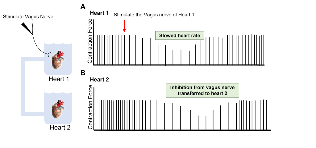
Acetylcholine was the first neurotransmitter discovered and chemically isolated, a feat which earned two researchers the shared Nobel Prize in Physiology or Medicine in 1936. One of the two scientists, a German pharmacologist named Otto Loewi, stimulated the vagus nerve connected to an isolated frog heart, which caused the heart rate to slow down. When he applied the surrounding solution from this experiment to a second heart, he observed that the second heart also slowed down, despite having no physical connection to the first heart. From this, he concluded that a chemical was being released by the vagus nerve that was decreasing the heart rate. This chemical was first called Vagusstoff, the German word meaning Vagus substance. Today, we know it as acetylcholine.
Acetylcholine Synthesis and Storage
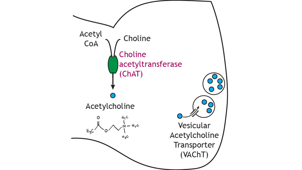
Acetylcholine (ACh) is a small molecule neurotransmitter best known for its role at the neuromuscular junction, the synapse between a motor neuron and a muscle fiber. In the presynaptic terminal, acetylcholine is synthesized from acetyl coenzyme A (acetyl CoA) and choline via the enzyme choline acetyltransferase (ChAT). The level of enzyme activity is the rate-limiting step in the synthesis pathway. The presence of ChAT in a neuron is used as a biochemical marker for neurons that produce acetylcholine. Acetylcholine is packaged into small vesicles for storage in the terminal via the vesicular acetylcholine transporter (VAChT), a protein found in the synaptic vesicle membrane.
Acetylcholine Receptors
Nicotinic Acetylcholine Receptors
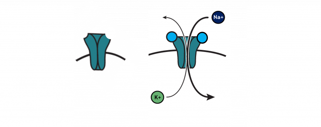
Acetylcholine is able to act at both ionotropic and metabotropic receptors, and activity at both receptor classes is essential for normal function. The ionotropic receptors of the nervous system are called nicotinic acetylcholine receptors because they can be activated by nicotine in addition to acetylcholine. Nicotinic receptors are located primarily outside of the central nervous system in the periphery. Specifically, the nicotinic receptors are used at the neuromuscular junction, which is the junction of a motor neuron and a skeletal muscle. Acetylcholine is released by motor neurons, where it activates nicotinic acetylcholine receptors on skeletal muscle cells. This excites the muscle cells and causes them to contract.
Nicotinic acetylcholine receptors are non-selective cation channels. The channel is closed when acetylcholine is not bound to the receptor. The nicotinic receptor has two binding sites for acetylcholine. When acetylcholine is bound to both sites on the receptor, the pore of the channel opens to allow movement of both sodium and potassium. This ionotropic receptor is an example of a direct mechanism of action, due to the fact that the receptor and the channel are located on the same protein. The properties of the receptors cause more sodium to enter than potassium leaves, ultimately causing the membrane to depolarize. Thus, nicotinic receptors are always excitatory (produce EPSPs) and cause depolarization of the postsynaptic cell.
Muscarinic Receptors
On the other hand, muscarinic acetylcholine receptors are GPCRs (or metabotropic receptors) and utilize an indirect mechanism of action. When acetylcholine binds to muscarinic receptors, the beta-gamma subunit of the G-protein complex translocates in the cell to affect potassium channels in the cell membrane, causing them to open. Due to the electrochemical gradient of potassium, potassium will leave the cell and cause membrane hyperpolarization, or IPSPs.
Muscarinic receptors are located in the heart, and their activation causes a decrease in heart rate due to this hyperpolarization caused by the muscarinic receptors (as Otto Loewi demonstrated with the isolated frog heart preparation.)
Animation 15.1. Some GPCRs, like the muscarinic acetylcholine receptors in the heart, alter cellular permeability by opening ion channels. The activated beta-gamma subunit of the muscarinic receptor opens GIRK potassium channels and allows the efflux of potassium. ‘Beta-Gamma Ion Channels’ by Casey Henley is licensed under a Creative Commons Attribution Non-Commercial Share-Alike (CC BY-NC-SA) 4.0 International License. View static image of animation.
Neuropharmacology and Acetylcholine Systems
Different drugs have the ability to interact within a neurotransmitter system, in many cases by directly interacting with a neurotransmitter receptor protein. There are two main classes of drugs that we will discuss: agonists and antagonists. An agonist is a drug that binds to a receptor and acts to activate the normal function of that receptor. An antagonist is a drug that binds to a receptor that acts to block the normal function of that receptor. The acetylcholine neurotransmitter system can be used as an example to see the function of both agonists and antagonists.
Drugs that interact with the nicotinic acetylcholine receptor
Nicotine is an agonist for the nicotinic acetylcholine receptor (which is how the receptor got its name). Normally, the nicotinic receptor causes excitatory postsynaptic potentials. Because nicotine is an agonist for this receptor, when it binds to the nicotinic acetylcholine receptor it also causes excitatory post synaptic potentials and lead to an overall increase in activity.
There are also antagonists for the nicotinic acetylcholine receptor. Curare, a substance that comes from the resin of a tree in South America and α-bungarotoxin, a toxin found in Krait snake venom, are both examples of antagonists for the nicotinic acetylcholine receptor. Recall that normally the nicotinic acetylcholine receptor causes excitatory post synaptic potentials. As antagonists, curare and α-bungarotoxin will block excitatory postsynaptic potentials, leading to an overall decrease in activity.
Drugs that interact with the muscarinic receptor
Muscarine, a substance found in certain mushrooms, is an agonist for the muscarinic receptor (and how the receptor got its name). As an agonist, muscarine will promote the normal function of the receptor. Normally, muscarinic receptors produce inhibitory post synaptic potentials. Thus, muscarine binding to muscarinic receptor will cause inhibitory post synaptic potentials, causing an overall decrease in activity.
Atropine is a drug that acts as an antagonist at the muscarinic receptor. Antagonists block the normal function of the receptor. Because normally the muscarinic receptor is inhibitory, atropine will block the normal inhibitory effects, leading to an overall increase in activity.
Termination of Acetylcholine Signaling
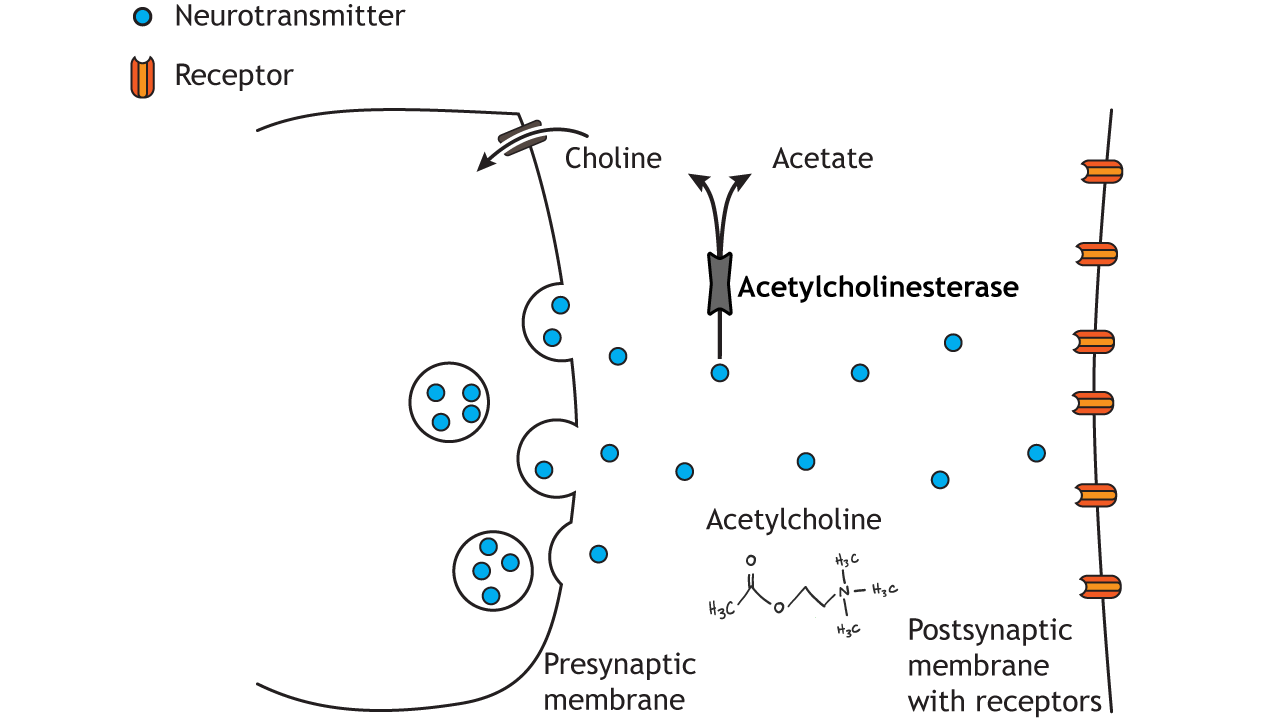
The enzyme acetylcholinesterase, located in the synaptic cleft and within the postsynaptic membrane, chemically breaks down acetylcholine into acetic acid and choline.
Choline can also undergo the process of reuptake which will remove choline from the synapse (which will effectively stop acetylcholine signaling) and bring it into the presynaptic cell through the choline transporter. Acetylcholinesterase is an important enzyme to regulate acetylcholine within the body. Recall that acetylcholine is used at the neuromuscular junction between motor neurons and skeletal muscles. The acetylcholinesterase enzyme prevents continuous stimulation of our muscles, allowing tight control. In fact, acetylcholinesterase is one of the fastest acting enzymes within the body. Nerve gas and neostigmine (in Nigerian beans) both act to inhibit acetylcholinesterase, causing excessive acetylcholine signaling.
Key Takeaways
- Acetylcholine was the first identified neurotransmitter and was discovered by Otto Loewi
- Acetylcholine is synthesized within the presynaptic terminal from choline and acetate through ChAT and packaged into vesicles by VAchT
- Acetylcholine binds to both ionotropic (nicotinic) and metabotropic (muscarinic) receptors
- Nicotinic receptors are excitatory and muscarinic receptors are inhibitory
- Agonists are drugs that promote the normal activity of the receptor that they bind to
- Antagonists are drugs that block the normal activity of the receptor that they bind to
- Acetylcholine signaling is terminated through the enzymatic activity of acetylcholinesterase
Test Yourself!
Attributions
Portions of this chapter were remixed and revised from the following sources:
- Foundations of Neuroscience by Casey Henley. The original work is licensed under a Creative Commons Attribution-NonCommercial-ShareAlike 4.0 International License
- Open Neuroscience Initiative by Austin Lim. The original work is licensed under a Creative Commons Attribution-NonCommercial 4.0 International License.
Media Attributions
- Acetylcholine Experiment © Valerie Hedges is licensed under a CC BY-NC-SA (Attribution NonCommercial ShareAlike) license
- Acetylcholine Synthesis © Casey Henley is licensed under a CC BY-SA (Attribution ShareAlike) license
- Nicotinic Receptor © Casey Henley adapted by Valerie Hedges is licensed under a CC BY-SA (Attribution ShareAlike) license
The synapse between a motor neuron and a muscle fiber
ionotropic receptors for acetylcholine
post-synaptic graded depolarization
When the membrane potential gets closer to zero
G-protein coupled receptors for acetylcholine
When the membrane potential gets further away from zero
a drug that binds to a receptor and acts to activate the normal function of that receptor
a drug that binds to a receptor that acts to block the normal function of that receptor
enzyme that breaks down acetylcholine
Process where neurotransmitters are removed from the synapse and transported back into the presynaptic cell to be re-packaged into synaptic vesicles
