
28 Spinal Control of Movement
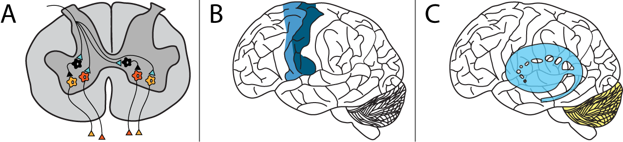
The motor system controls all of our skeletal muscle movement. There are multiple levels of control. Within the spinal cord, simple reflexes can function without higher input from the brain. Slightly more complex spinal control occurs when central pattern generators function during repetitive movements like walking. The motor and premotor cortices in the brain are responsible for the planning and execution of voluntary movements. And finally, the basal ganglia and cerebellum modulate the responses of the neurons in the motor cortex to help with coordination, motor learning, and balance.
This lesson explores the lowest level of control – spinal reflexes.
Alpha Motor Neurons
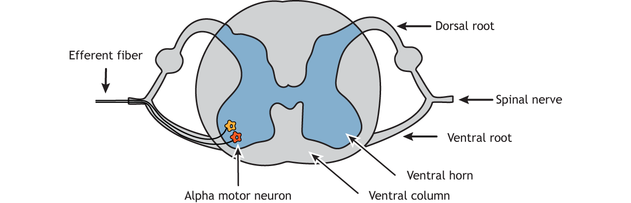
Muscle fibers are innervated by alpha motor neurons. The cell bodies of the alpha motor neurons are located in the central nervous system in the ventral horn of the spinal cord. Their axons leave the spinal cord via the ventral roots and travel to the muscle via efferent peripheral spinal nerves.
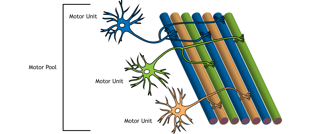
One alpha motor neuron can innervate multiple fibers within one muscle; the axons of a motor neuron can branch to make synaptic contacts with many fibers. A motor neuron and the fibers innervated by it are called a motor unit. The muscle fibers within one motor unit are often spread throughout the muscle to spread the contraction throughout the full muscle.
The group of motor neurons that innervate all the fibers of one muscle is called a motor pool.
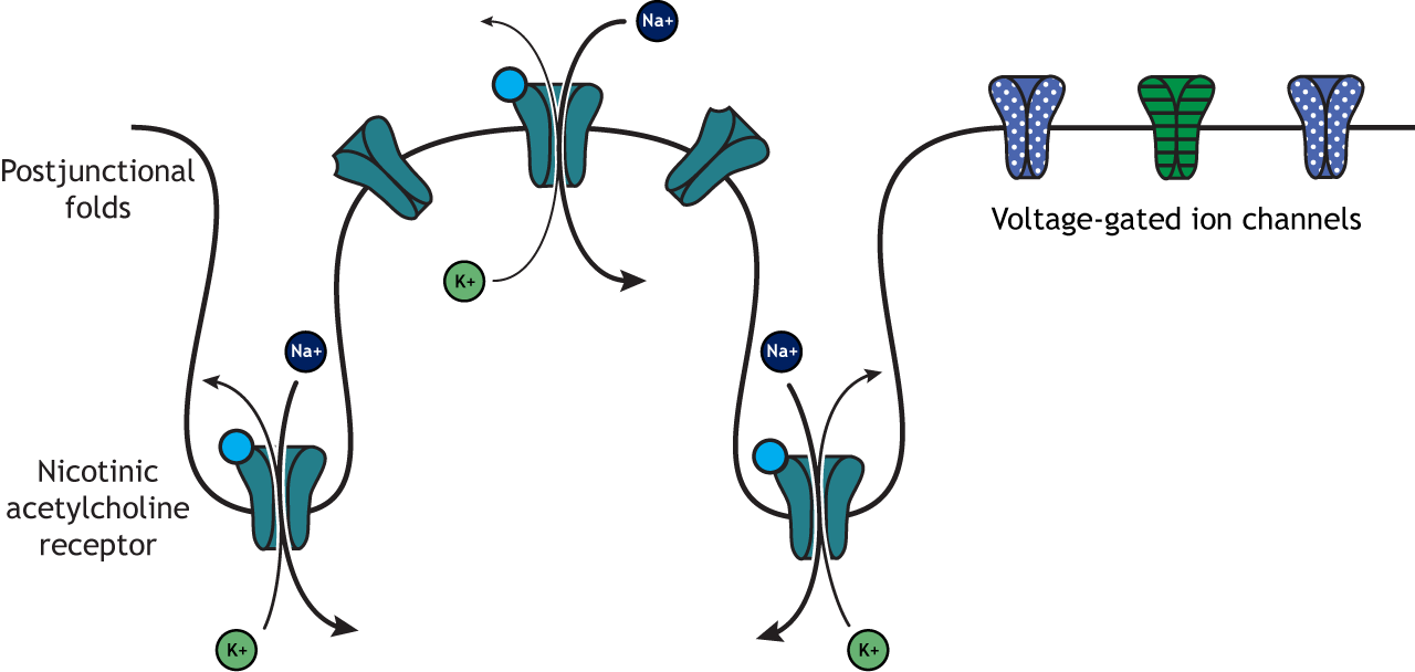
Neuromuscular Junction
The neuromuscular junction is one of the largest synapses in the body and one of the most well-studied because of its peripheral location. Acetylcholine is the neurotransmitter released at the neuromuscular junction (NMJ), and it acts upon ligand-gated, non-selective cation channels called nicotinic acetylcholine receptors that are present in postjunctional folds of the muscle fiber. Acetylcholinesterase, an enzyme that breaks down acetylcholine and terminates its action, is present in the synaptic cleft of the neuromuscular junction.
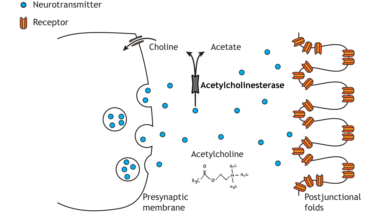
Nicotinic acetylcholine receptors allow the influx of sodium ions into the muscle cell. The depolarization will cause nearby voltage-gated channels to open and fire an action potential in the muscle fiber. In a healthy system, an action potential in the motor neurons always causes an action potential in the muscle cell. The action potential leads to contraction of the muscle fiber.
Organization
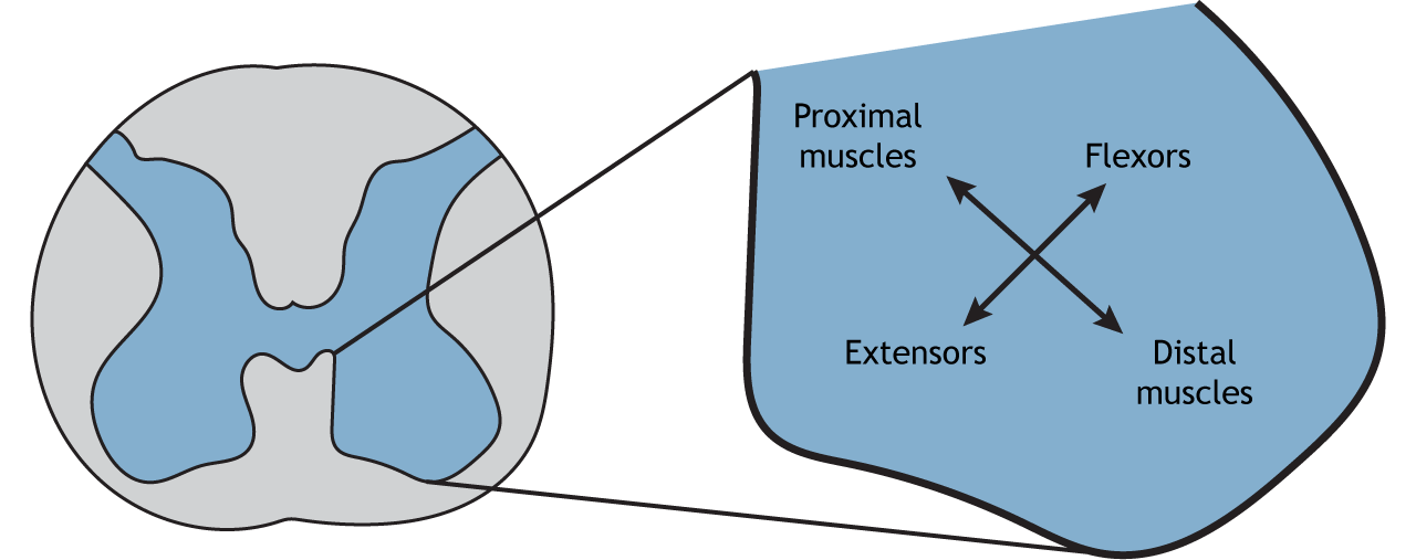
Like the sensory systems, the motor system is also organized in a topographic fashion. Within the spinal cord, alpha motor neurons that innervate muscles in the arms and legs are located in the lateral portion of the ventral horn, whereas alpha motor neurons that innervate muscles in the trunk are located in the medial portion.
Sensation
Proprioception is the ability to know where your body is in space and relies on the presence of sensory receptors located within the muscles. Some of these specialized structures are called muscle spindles, and they monitor muscle fiber stretch. Information is relayed to the nervous system via Group I sensory axons, which are large, myelinated fibers. Muscle spindles are important for spinal reflexes.

Reflexes
Stretch (Myotatic) Reflex
The stretch reflex, also called the myotatic, patellar, or knee-jerk reflex, occurs in response to activation of the muscle spindle stretch receptors. The stretch reflex is a common occurrence at a doctor’s visit when the doctor taps your knee with a little hammer. This usually results in the lower leg kicking up slightly. The synaptic communication for this reflex takes place completely within the spinal cord and requires no input from the brain.
The knee is tapped on the tendon that connects to the quadriceps muscle. The tendon extends enough to stretch the quadriceps muscle, activating the stretch receptors. Sensory information travels to the dorsal horn of the spinal cord where it synapses on alpha motor neurons that innervate the quadriceps. Activation of the motor neurons contracts the quadriceps, extending the lower leg. This is called monosynaptic communication because there is only one synapse between the sensory input and the motor output.
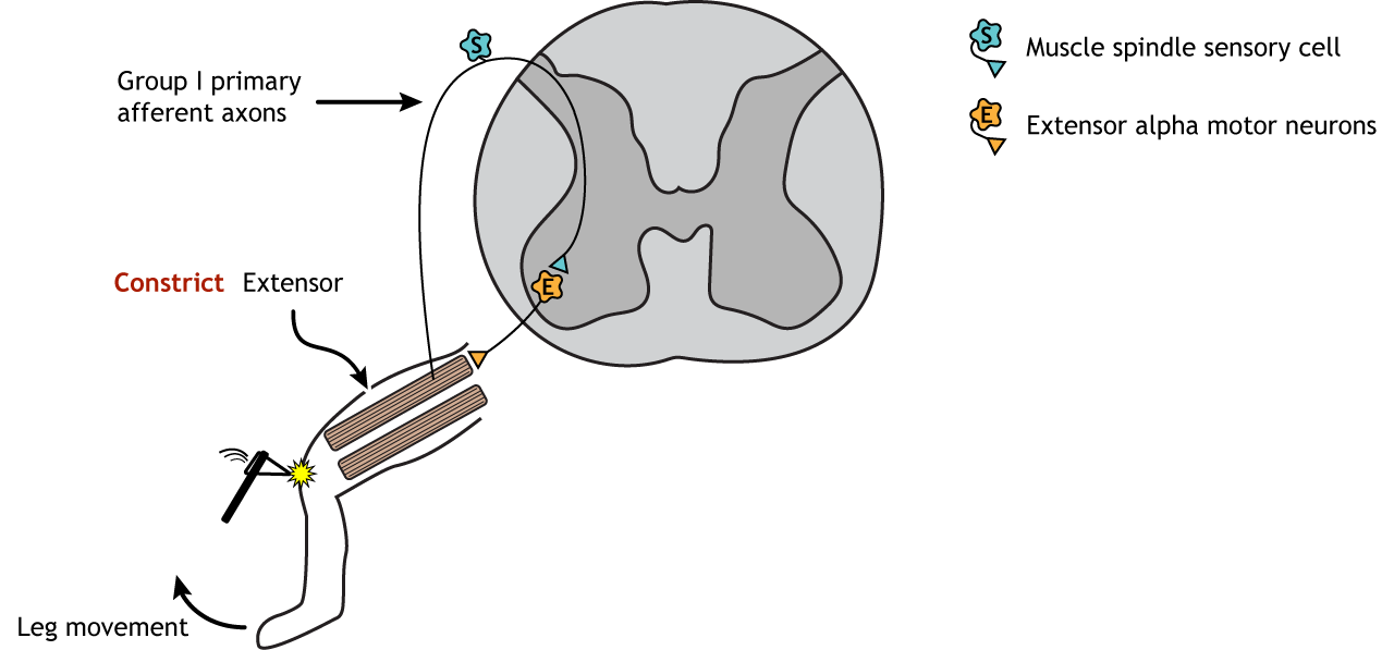
The sensory neurons also synapse on interneurons within the spinal cord that are inhibitory. These inhibitory interneurons then synapse on alpha motor neurons that innervate the hamstring, the antagonistic flexor muscle to the quadriceps. When these motor neurons are inhibited, the hamstring muscle relaxes, allowing the contraction of the quadriceps to occur with more ease.

Withdrawal (Flexor) Reflex
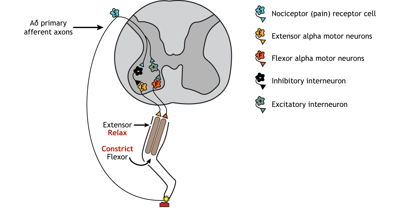
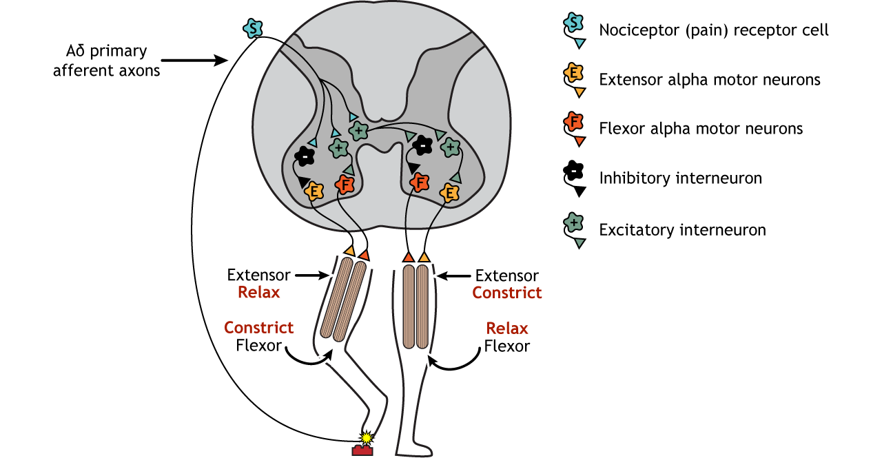
A similar process can be seen in the withdrawal reflex. In this case, instead of an extension, the muscles lead to muscle flexion in response to a stimulus. If, for example, you step on something painful, the reflex will be to lift the injured foot. The sensory information that initiates this reflex is activation of pain receptors, or nociceptors. Like with the stretch reflex, the sensory information enters the spinal cord at the dorsal horn. Unlike the stretch reflex, the withdrawal reflex is a polysynaptic reflex, meaning interneurons are present between the sensory neurons and the motor neurons. Excitatory interneurons communicate with the alpha motor neurons of the flexor muscle, whereas inhibitory interneurons communicate with the alpha motor neurons of the extensor muscle. The behavioral response is flexing of the leg upward (the opposite action of the stretch reflex).
Crossed-Extensor Reflex
Running in parallel to the withdrawal reflex is the crossed-extensor reflex. If you step on something sharp and lift that leg, your other leg needs to be able to support your weight shift, or you would fall. This is accomplished by interneurons that cross the midline of the spinal cord and communicate with motor neurons on the contralateral side of the body. The painful sensory information that initiated the withdrawal reflex also initiates the crossed-extensor reflex. In addition to the ipsilateral interneurons active in the withdrawal reflex, the sensory axons also synapse on excitatory interneurons that cross the midline. These interneurons then synapse on excitatory interneurons that activate the alpha motor neurons of the extensor muscle and inhibitory interneurons that inhibit the alpha motor neurons of the flexor muscle (the opposite configuration to the withdrawal reflex). This leads to the leg extending, providing a stable base for the weight shift.
Central Pattern Generators
Locomotion
Locomotion is one example of a basic, rhythmic movement that requires coordination of a number of muscle groups to work properly (other examples include swimming, flying, respiration, swallowing).
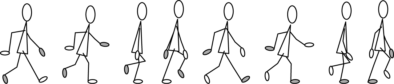
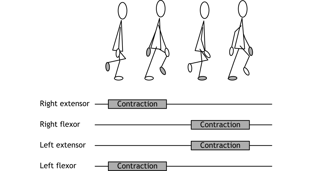
Activity of extensor and flexor muscles in both legs must be coordinated to allow smooth locomotion without falling. These rhythmical movements are controlled at the level of the spinal cord by circuits called central pattern generators. The spinal cord has circuitry that, in the case of walking, moves the legs in opposite patterns. When one leg is lifting up to move forward, the other leg is stable, touching the ground.
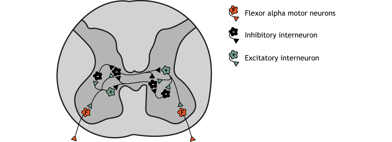
Spinal Circuitry
The control of this system has multiple levels. Neurons themselves may have pacemaker properties that allow for a continuous cycle of depolarization and repolarization. These neurons are then located within multi-cell circuits involving a collection of excitatory and inhibitory interneurons that results in reciprocal inhibition of contralateral muscles. Additional networks of spinal interneurons would cause reciprocal inhibition of ipsilateral antagonistic muscles.
Although the spinal cord is able to control these movements on its own, there is input from both the brainstem and sensory neurons which can have an effect on modulating the pattern of neuronal activity in the spinal cord. For example, when an animal needs to slow down, speed up, or turn away from a danger, for example, those inputs can alter the spinal cord circuit.
Key Takeaways
- Motor neuron cell bodies are located in the ventral horn of the spinal cord
- Motor neuron axons are located in the peripheral nervous system and travel to muscles via spinal nerves
- Acetylcholine is released at the neuromuscular junction and acts upon ionotropic nicotinic acetylcholine receptors
- The spinal cord is topographically organized
- Control of reflexes occurs within the spinal cord and input from the brain is not needed
- Central pattern generators are circuits in the spinal cord that control repetitive, consistent movements like walking
Test Yourself!
- What is the difference between a motor unit and a motor pool?
Video Lecture
