
18 Internal Brain Anatomy
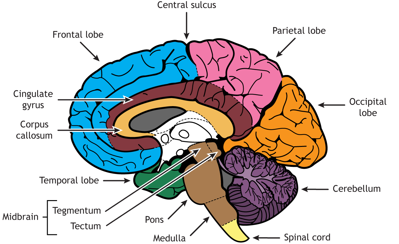
A mid-sagittal section slices the brain through the longitudinal fissure and separates the right hemisphere from the left. It also reveals more structures. In a mid-sagittal view, all four cortical lobes are visible. The frontal lobe is separated from the parietal lobe by the central sulcus, the occipital lobe is in the posterior region of the brain, and the temporal lobe can be seen behind the brainstem. The cerebellum, pons, medulla, and spinal cord are seen caudal to the cerebrum, but in this view, the midbrain, which is made up of two regions, the tegmentum and tectum, are also visible superior to the pons. The corpus callosum is located in the center of the cerebrum and is a white matter bundle made up of axons crossing from one hemisphere to the other. Surrounding the corpus callosum is the cingulate gyrus, a region important for emotion.
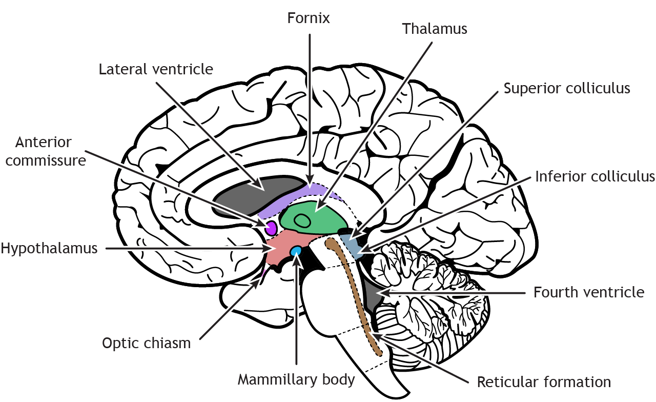
The diencephalon consists of subcortical structures and connects the forebrain to the midbrain
The diencephalon region of the brain consists of the region around the thalamus and hypothalamus. It is located inferior to the fornix and lateral ventricle, posterior to the anterior commissure, and superior to the brainstem. The fornix is a nerve fiber bundle containing primarily output from the hippocampus. The anterior commissure sits above the hypothalamus and is white matter tract, like the corpus callosum, that allows information to cross from one hemisphere to the other. The thalamus is best known for its role as a relay and processing location for the sensory and motor systems. The hypothalamus has a variety of functions including control of stress and the “fight or flight” response of the autonomic nervous system, reproduction, sleep, thirst, hunger, and other homeostatic functions. The mamillary bodies sit in the posterior part of the hypothalamus and are important for memory. The optic nerves from the retina cross at the optic chiasm, and then the optic tracts continue back into the diencephalon.
In the brainstem, the tectum of the midbrain consists of the superior and inferior colliculi, which are important for vision and hearing, respectively. The reticular formation is located throughout the brainstem. Networks within the reticular formation are important for regulating sleep and consciousness, pain, and motor control. The fourth ventricle lies between the brainstem and the cerebellum.
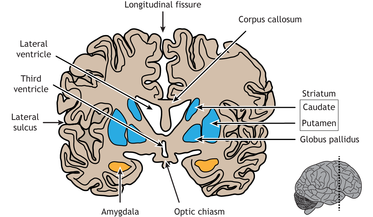
The basal ganglia, amygdala, and hippocampus are subcortical forebrain structures with a range of functions
Coronal sections of the brain allow deep tissue structures to be visible. A cut through the anterior portion of the temporal lobe shows the amygdala, a region important for emotion, located in the medial temporal lobe. The regions of the basal ganglia are also visible; the striatum, which consists of the caudate and the putamen, and the globus pallidus. The basal ganglia has multiple functions but is best known for its role in regulation of movement. The lateral ventricle sits medial to the basal ganglia, and above the lateral ventricle is the corpus callosum. The third ventricle is located in the middle of the brain, inferior to the lateral ventricle, and the optic chiasm lies inferior to the third ventricle. The longitudinal fissure separates the left and right cerebral hemispheres, and the lateral sulcus is the border between the frontal and temporal lobes.
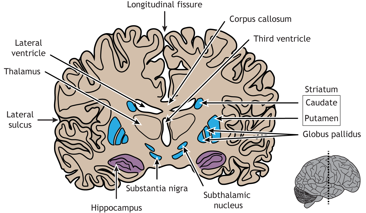
A coronal section taken closer to the central sulcus will make the hippocampus visible. The hippocampus is known for its role in memory and spatial awareness. At this location, the basal ganglia is more defined; the caudate and putamen are still present, but the two separate regions of the globus pallidus, the internal and external segments, can be seen, as well as the subthalamic nucleus and the substantia nigra. The thalamus is located on either side of the third ventricle. The corpus callosum is superior to the lateral ventricle. The cerebrum is divided in half by the longitudinal fissure, and the lateral sulcus separates the temporal lobe from the frontal and parietal lobes.
Key Takeaways
- Like the external anatomy, internal structures also have specific functions
- Sectioning the brain in different ways provides views of subcortical regions
Test Yourself!
