
24 Touch: The Skin
Touch can come in many forms: pressure, vibration, stretch, motion, edges, points, etc. Receptors in the skin allow for perception of these different characteristics, and when this information is combined in the central nervous system, we are able to determine the location, strength, duration, movement, shape, and texture of the object interacting with the skin.
Receptors
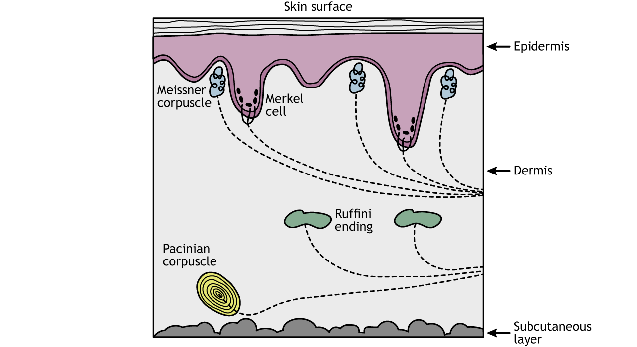
We can feel different modalities of touch because of the presence of specialized sensory receptors, called mechanoreceptors, located in the skin.
The Pacinian corpuscles are located deep in the dermis of the skin and are responsible for perception of vibration.
Ruffini endings detect skin stretch and are also located within the dermis layer of the skin.
The Meissner corpuscles are stimulated by skin motion and are located in the epidermis layer.
The Merkel cells are located at the border between the dermis and epidermis and are specialized to detect edges and points.
Multiple types of mechanoreceptors allow for perception of different qualities of touch
Receptive Fields
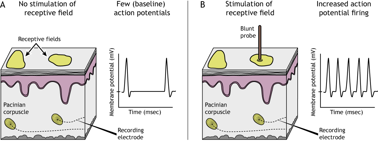
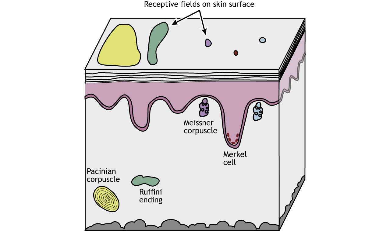
Each mechanoreceptor responds to a touch stimulus in a specific area of the skin, a region called the receptive field of the receptor. When the receptive field is touched, the mechanoreceptor will be activated.
Receptive field characteristics differ depending on the type of mechanoreceptor and location on the body
Receptive Field Size
Merkel cells and Meissner corpuscles, both of which are located near the skin surface, have small receptive fields. Ruffini endings and Pacinian corpuscles, located deeper in the skin layers, have larger receptive fields than the Merkel cells and Meissner corpuscles.
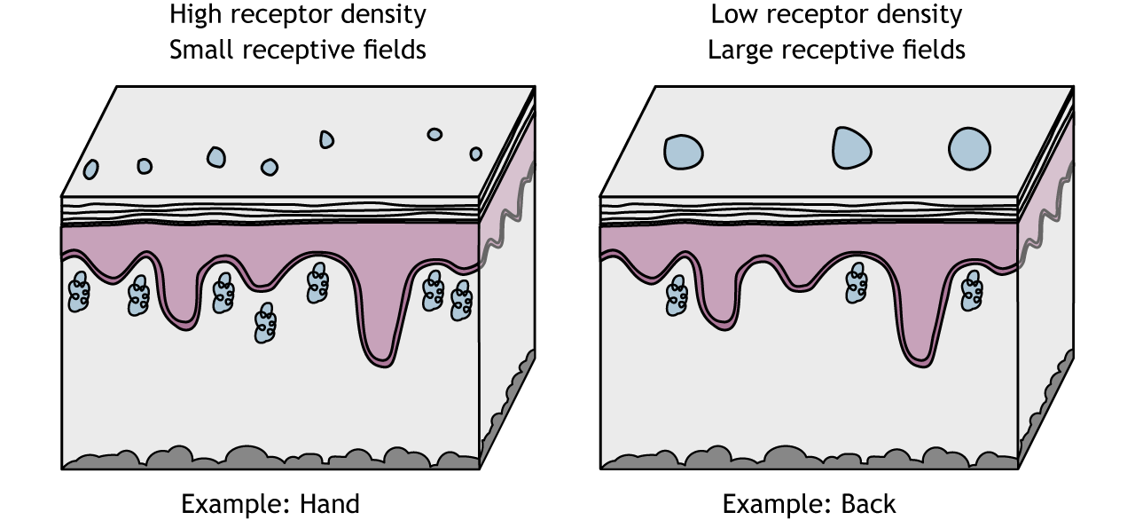
Receptive field sizes are different among the different mechanoreceptors, but they also vary among different body regions. Even within one receptor type (e.g. Meissner corpuscles), receptive fields in regions like the fingers or lips are smaller than in regions like the back or leg. This allows us to have finer spatial resolution with locating and identifying objects using our fingers. The smaller receptive fields in these regions are a result of a higher density of receptors in the skin.
Two-Point Discrimination
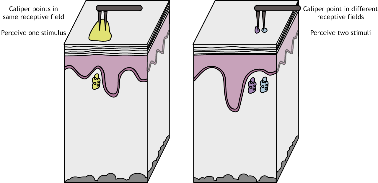
Receptive field sizes are important because they allow us to locate a stimulus on our bodies. Larger receptive fields are not as precise as smaller receptive fields. One measure of receptive field size is two-point discrimination (try it at home!), which determines the minimum distance needed between two stimuli to perceive two separate points on the skin and not one. The hand has a smaller threshold for discerning between two points than does the back, a result of the different sized in receptive fields.
Adaptation Rate
Another important characteristic of the somatic sensory receptors is that of adaptation rate. Fibers that are slowly adapting show action potential firing throughout the entire time a stimuli is present. Merkel cells and Ruffini endings are both slowly adapting fibers. Slowly adapting fibers are most useful for determining the pressure and shape of a stimulus.
Animation 24.1. Slowly adapting mechanoreceptors continuing firing action potentials throughout the duration of a stimulus. As the stimulus moves from not present, to weak, to strong, the action potential firing of the Ruffini ending fires throughout the entire stimulus. ‘Slowly Adapting Receptor’ by Casey Henley is licensed under a Creative Commons Attribution Non-Commercial Share-Alike (CC BY-NC-SA) 4.0 International License. View static image of animation.
Rapidly adapting fibers fire action potentials when a stimulus changes (e.g., starts, stops, gets stronger or weaker) but not when a stimulus is constant. This firing makes rapidly adapting fibers specialized for detecting movement and vibration. Meissner and Pacinian corpuscles are rapidly adapting.
Animation 24.2. Rapidly adapting mechanoreceptors firing action potentials when the strength of the stimulus changes. As the stimulus moves from not present, to weak, to strong, the action potential firing of the Pacinian corpuscle only fires when the stimulus changes strength. ‘Rapidly Adapting Receptor’ by Casey Henley is licensed under a Creative Commons Attribution Non-Commercial Share-Alike (CC BY-NC-SA) 4.0 International License. View static image of animation.
Sensory Transduction
In previous chapters we discussed ion channels that are gated by voltage changes in the neuron and channels that are gated by neurotransmitters. In the somatosensory system, we find ion channels that are gated by physical distortion or stretch of the membrane. These channels can open by stretch of the membrane itself or indirectly through movement of intra- or extracellular proteins that are linked to the channels. Sodium and calcium flow into the cell, causing both a depolarization and the initiation of second messenger cascades. If enough stimulus is applied, the depolarization reaches threshold of the axon and an action potential is sent toward the spinal cord.
Animation 24.3. Mechanoreceptors respond to touch stimuli via stretch-gated non-selective cation channels. The channels can either open due to stretch of the membrane itself which stretches open the channel or due to proteins associated with the channels that pull the channel open. ‘Stretch-Gated Ion Channels’ by Casey Henley is licensed under a Creative Commons Attribution Non-Commercial Share-Alike (CC BY-NC-SA) 4.0 International License. View static image of animation.
Key Takeaways
- There are multiple types of mechanoreceptors in the skin that are activated by different types of touch stimuli
- The receptive field size differs among the types of mechanoreceptors
- The adaptation rate differs among the types of mechanoreceptors
- Receptive field is a region of skin that activate a given mechanoreceptor
- Receptive field size for a specific type of mechanoreceptor can vary in size across the body
- Mechanoreceptors express stretch-gated non-selective ion channels that depolarize the cell during sensory transduction
Test Yourself!
- Describe the relationship between density of receptors, receptive fields, and two-point discrimination.
