16 Anatomical Terminology

Familiarity with the terminology used to describe location and relationships within the nervous system is critical as we move forward into examining brain systems.
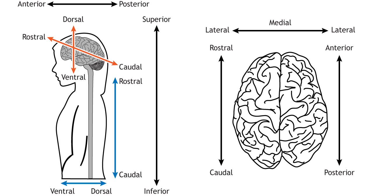
Directional Terms

Directional terms are used to locate one structure, usually in relation to another structure. Some terms, like dorsal or ventral, are relative to the axis of the central nervous system, so the direction these terms define changes if used for brain regions versus other body regions. Other terms, like superior or inferior, keep their meaning across the entire body.

  • Anterior: In front of; toward the face
  • Posterior: Behind; toward the back
  • Superior: Above; toward the head
  • Inferior: Below; toward the feet
  • Medial: Toward the middle
  • Lateral: Toward the edge
  • Dorsal: Toward the top of the brain or the back of the spinal cord
  • Ventral: Toward the bottom of the brain or the front of the spinal cord
  • Rostral: Toward the front of the brain or the top of the spinal cord
  • Caudal: Toward the back of the brain or the bottom of the spinal cord
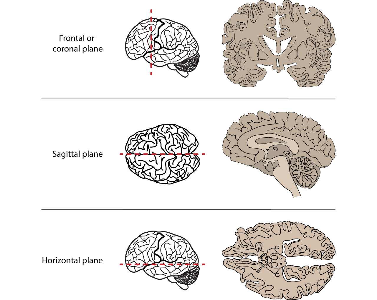
Illustration of a body and a brain showing directional anatomical terms. Details in caption and text.
Figure 16.1. Directional terms used to locate nervous system structures. The dorsal / ventral and rostral / caudal pairs point in different directions depending on if they are referring to the axis of the brain (orange arrows) or the axis of the spinal cord (blue arrows). The definitions of each term are described in the text. ‘Anatomical Directions’ by Casey Henley is licensed under a Creative Commons Attribution Non-Commercial Share-Alike (CC BY-NC-SA) 4.0 International License.

Anatomical Planes

There are planes or axes that can be used to examine the nervous system. The frontal or coronal plane is a vertical plane in a medial to lateral direction, dividing objects into front and back pieces. The sagittal plane is also a vertical plane but in a rostral-caudal direction, meaning it divides objects into right and left regions. Finally, the horizontal plane divides objects into top and bottom regions.

Illustrations of anatomical planes and brain slices created by those planes. Details in caption.
Figure 16.2. Three anatomical planes are used to divide the nervous system to be able to view internal regions and structures. The frontal or coronal plane is a vertical plane that runs parallel to the eyes or ears and will divide the body into front and back regions. The sagittal plane is a vertical plane that runs perpendicular to the eyes or ears and will divide the body into left and right regions. The horizontal plane runs parallel to the ground and will divide the body into top and bottom regions. ‘Anatomical Planes’ by Casey Henley is licensed under a Creative Commons Attribution Non-Commercial Share-Alike (CC BY-NC-SA) 4.0 International License.

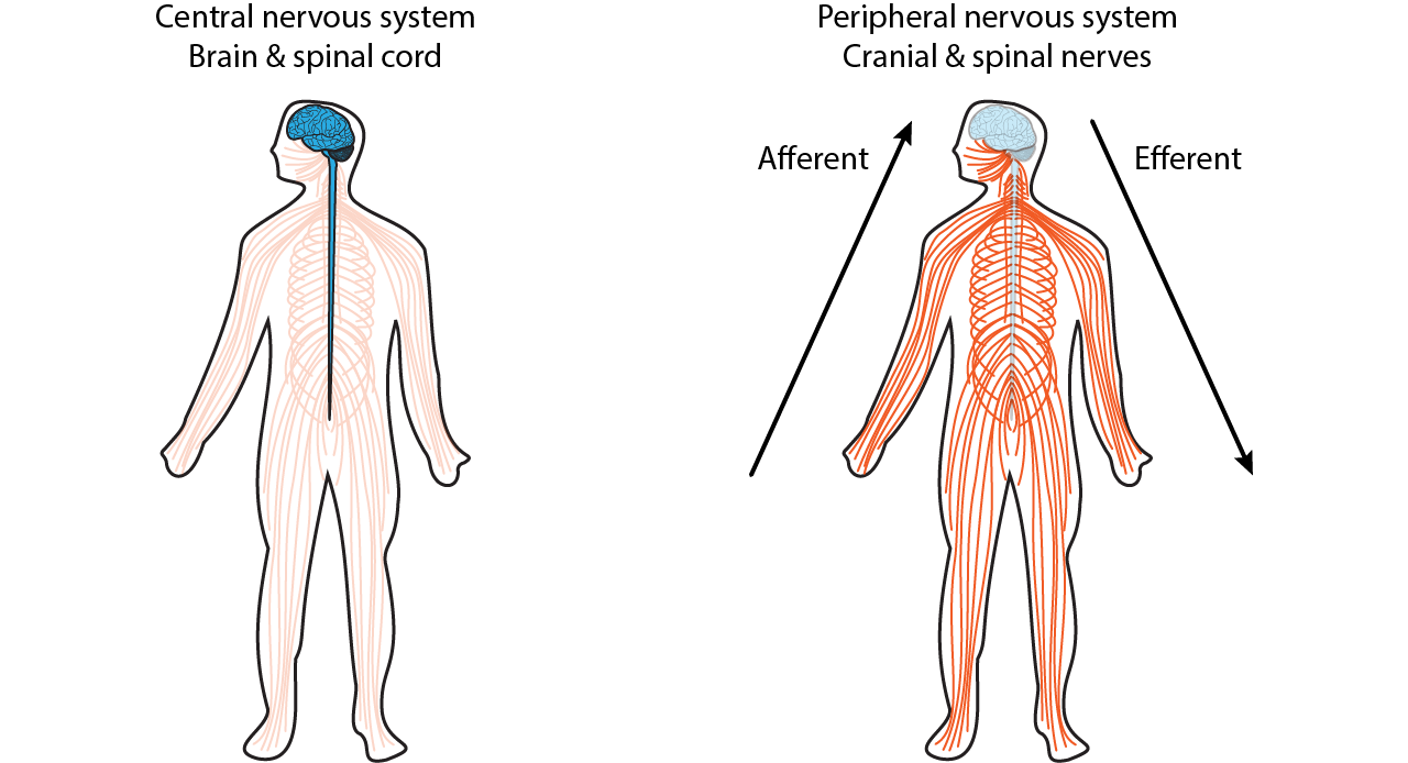
Nervous System Divisions

The nervous system is divided into two primary components. The central nervous system (CNS) is comprised of the brain and the spinal cord. The peripheral nervous system (PNS) is comprised of the cranial and spinal nerves. When information flow is described in the nervous system, it can either be afferent communication, meaning it is moving from the periphery to the brain, or efferent communication, meaning it is moving from the brain to the periphery.

Illustration of two bodies showing the central and peripheral nervous systems. Details in caption.
Figure 16.3. The nervous system is divided into the central nervous system, which includes the brain and spinal cord, and the peripheral nervous system, which includes the cranial and spinal nerves. Information traveling toward the brain is called afferent, whereas information traveling from the brain is called efferent. ‘CNS and PNS’ by Casey Henley is licensed under a Creative Commons Attribution Non-Commercial Share-Alike (CC BY-NC-SA) 4.0 International License.

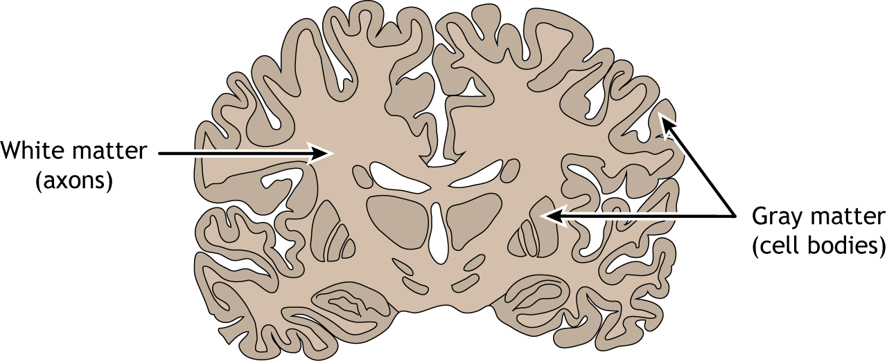
Tissue in the central nervous system can be further divided into either white matter or gray matter. White matter regions are comprised of axons. It appears white due to the myelin sheath on the axons. Gray matter regions are comprised of cell bodies and dendrites. Gray matter is the location of most synapses.

Illustration of a frontal brain section showing white and gray matter. Details in caption.
Figure 16.4. The central nervous system tissue can be divided into white and gray matter. White matter is primarily myelinated axons. Gray matter is primarily neuronal cell bodies and dendrites. In the brain, the surface of the cerebral cortex is a layer of gray matter. White matter can be found below the gray matter layer and is the location of the axons traveling to and from the cortical cell layer. Gray matter can also be found deep in the brain in subcortical regions that play critical roles in behavior. ‘White and Gray Matter’ by Casey Henley is licensed under a Creative Commons Attribution Non-Commercial Share-Alike (CC BY-NC-SA) 4.0 International License.

Key Takeaways

  • Anatomical terminology is critical for determining neurological landmarks

Test Yourself!

Video Lecture

License

Icon for the Creative Commons Attribution-NonCommercial-ShareAlike 4.0 International License

Foundations of Neuroscience Copyright © 2021 by Casey Henley is licensed under a Creative Commons Attribution-NonCommercial-ShareAlike 4.0 International License, except where otherwise noted.

Share This Book