
23 Somatosensory Systems
The somatosensory system is regulated by receptors that are spread throughout the body and measure a number of different sensory modalities in the body. These sensations can be divided into three main divisions: external stimuli, internal stimuli, and the sense of where the body is in space.
Perception of external stimuli can include the sense of touch (via mechanoreceptors), pain (via nociceptors), and temperature (via thermal receptors). Perception of internal stimuli can include organ (visceral) sensation and pain (via multiple receptor types) and blood chemical composition (via chemoreceptors). Finally, proprioception (via proprioceptors) is the sense of where the body is in space. The ability of an individual to touch their nose easily while their eyes are closed is an example of the proprioception system.
Somatosensory Cell Bodies
All somatosensory receptor neurons have their cell bodies located in the dorsal root ganglion, a structure found just outside the dorsal aspect of the spinal cord. The receptor neurons, also called primary afferent fibers, of the somatosensory system are bipolar neurons, meaning they have one process from the cell body that splits into two branches. One travels to the location of the receptor (e.g. the skin for touch) via the spinal nerves, and one travels into the spinal cord at the dorsal horn via the dorsal root. The axon can either synapse locally in the spinal cord or ascend to the brain via the dorsal column.

Primary Afferent Axons
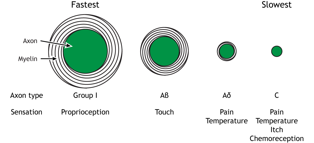
Primary afferent axons are divided into four groups based on size and conduction speed. The groups, unfortunately, have different names depending on if the axons come from the skin (Aα, Aβ, Aδ, and C fibers; examples are touch or pain) or the muscles (Group I, II, III and IV fibers; example is proprioception). The fastest axons are the Aα or Group I type; they have the largest diameter and are heavily myelinated. The next fastest myelinated axons are the Aβ or Group II fibers, followed by the Aδ or Group III fibers. Finally, the C fibers have the smallest diameter, are unmyelinated, and are the slowest at conducting action potentials.
| Afferent axon from muscle | Group 1 | Group II | Group III | Group IV |
| Afferent axon from skin | Aα | Aβ | Aδ | C |
| Diameter (µm) | 13-20 (Largest) | 6-12 | 1-5 | 0.2-1.5 (Smallest) |
| Conduction speed (m/sec) | 80-120 (Fastest) | 35-75 | 5-30 | 0.5-2 (Slowest) |
Table 21.1. Diameter and conduction speed of primary afferent axons. Group I and A alpha have the largest diameter and fastest speed. Group II and A beta are the next largest and fastest, followed by Group III and A delta. Group IV and C fibers are the slowest and smallest of all axon types.
Different sensory information is sent via the different types of axons. Proprioceptive information from the skeletal muscles is sent to the spinal cord via Group I fibers. Touch information from the mechanoreceptors travels along Aβ fibers. Aδ fibers carry pain and temperature sensation, and C fibers convey information about pain, temperature, itch, and chemoreception.
Dermatomes
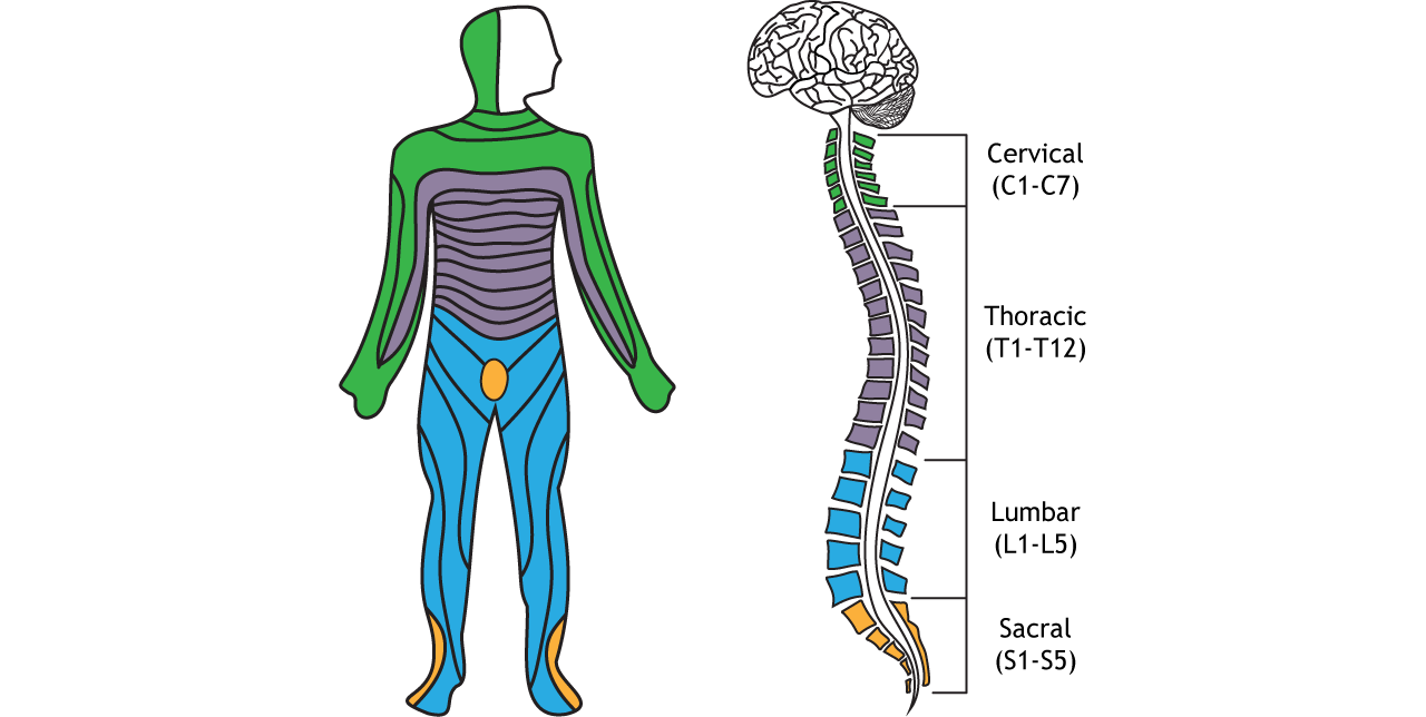
The afferent axons from the dorsal root ganglion enter the spinal cord via the spinal nerves. Axons from nearby regions of the body enter the spinal cord together, and this forms regions of skin that are innervated by the same spinal nerve. These regions are called dermatomes. Damage to a spinal nerve will cause dysfunction along the innervated dermatome. The dermatomes and spinal nerves are divided into 4 groups. The seven cervical spinal segments are the most rostral and are located in the neck. The twelve thoracic spinal segments are located along the chest and abdomen. The five lumbar segments are located below the thoracic segments, and the five sacral segments are the most caudal.
Key Takeaways
- Somatosensory neuron cell bodies are located in the dorsal root ganglion
- Somatosensory primary afferent axons ascend to the brainstem via the dorsal column white matter tract
- Primary afferent axons vary in diameter and myelination, both of which affect action potential speed
- Different somatosensory information is carried by the different sizes afferents
- Dermatomes are the region of skin innervated by one spinal nerve
Test Yourself!
