
29 Planning of Movement
There are a number of steps that must take place for voluntary movement to occur. Assessment of the surrounding environment and the body’s location in space, followed by determining what action is appropriate, and then initiating that action. We will first focus on the cortical regions involved in planning of voluntary movement.
After work, you sit down on the couch to watch one episode of your favorite show. As the end credits appear, you realize it is now time to head to your study space and start working on class. To do this, you need to leave the couch, grab your computer from the table, get your coffee from the kitchen and head to a different room. All of these voluntary movements take a great deal of processing by the brain. You must assess your surrounding environment and your body’s location in it, determine which actions need to be completed, and then actually initiate those actions. In this chapter we will focus on how the planning of voluntary movement occurs.
Cortical Anatomy
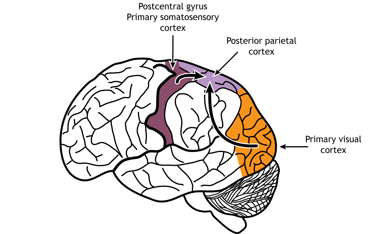
Much of the cortex is actually involved in the planning of voluntary movement. Sensory information, particularly the dorsal stream of the visual and somatosensory pathways, are processed in the posterior parietal lobe where Visual, tactile, and proprioceptive information are integrated.
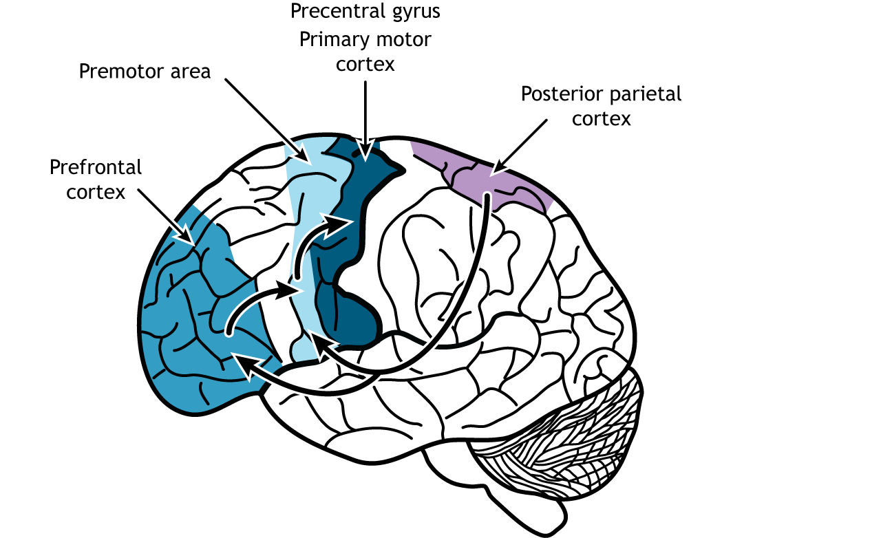
Connections from the posterior parietal lobe are then sent to both the premotor regions and the prefrontal cortex. The prefrontal cortex, which is located in the front of the brain in the frontal lobe, plays an important role in higher level cognitive functions like planning, critical thinking, and understanding the consequences of our behaviors. The premotor area lies just anterior to the primary motor cortex. This region helps plan and organize movement and makes decisions about which actions should be used for a situation.
View the primary motor cortex using the BrainFacts.org 3D Brain
View the premotor cortex using the BrainFacts.org 3D Brain
View the prefrontal cortex using the BrainFacts.org 3D Brain
Role of Premotor Area
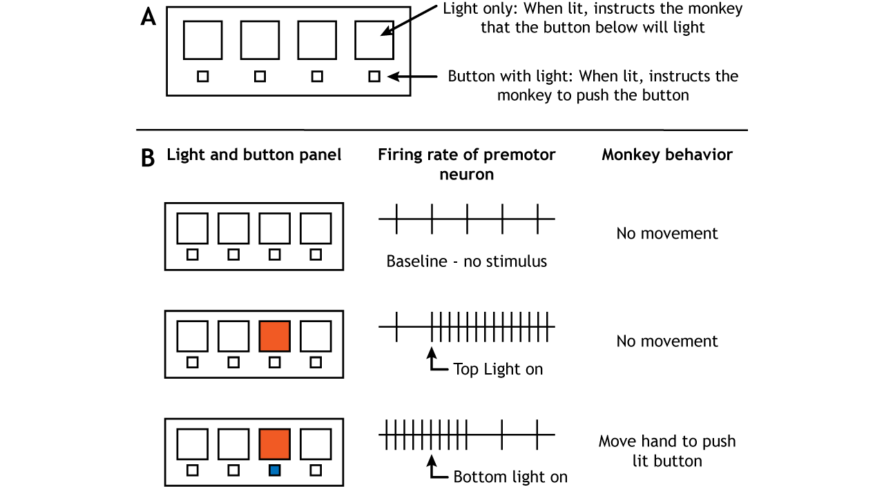
The premotor regions do send some axons directly to lower motor neurons in the spinal cord using the same pathways as the motor cortex (see Execution of Movement chapter). However, the premotor cortex also plays an important role in the planning of movement. Two experimental designs have demonstrated this role. Monkeys were trained on a panel that had one set of lights in a row on top and one set of buttons that could also light up in a row on the bottom. The monkeys would watch for a top row light to turn on. This would indicate that within a few seconds, the button directly below would light up. When the button turned on, the monkeys were supposed to push the button.
Therefore, there were two light triggers in the experiment. The first required no motor movement from the monkey but did give the monkey information about where a motor movement would be needed in the near future. The second required the monkey to move to push the button. When brain activity was measured during this study, neurons in the premotor cortex became active when the first light trigger turned on, well before any movement actually took place (Weinrich and Wise, 1928).
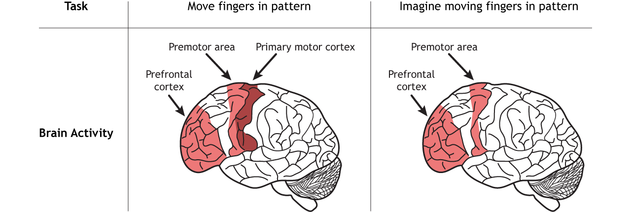
In another experiment, people were trained to move their fingers in a specific pattern. Cerebral blood flow was then measured when they repeated the finger pattern and when they only imagined repeating the finger pattern. When the movement was only imagined and not actually executed, the premotor regions along with parts of the prefrontal cortex were activated (Roland, et al, 1980).
These studies show that the premotor cortex is active prior to the execution of movement, indicating that it plays an important role in the planning of movement. The posterior parietal, prefrontal, and premotor regions, though, also communicate with a subcortical region called the basal ganglia to fully construct the movement plan. The basal ganglia are covered in the next chapter.
Key Takeaways
- Sensory information is processed in the posterior parietal before being sent to motor regions of the brain
- The prefrontal cortex and premotor cortex are critical for creating a movement plan
Test Yourself!
Video Lecture
References
Roland PE, Larsen B, Lassen NA, Skinhøj E. Supplementary motor area and other cortical areas in organization of voluntary movements in man. J Neurophysiol. 1980 Jan;43(1):118-36. doi: 10.1152/jn.1980.43.1.118. PMID: 7351547.
Weinrich M, Wise SP. The premotor cortex of the monkey. J Neurosci. 1982 Sep;2(9):1329-45. doi: 10.1523/JNEUROSCI.02-09-01329.1982. PMID: 7119878; PMCID: PMC6564318.
