
17 External Brain Anatomy
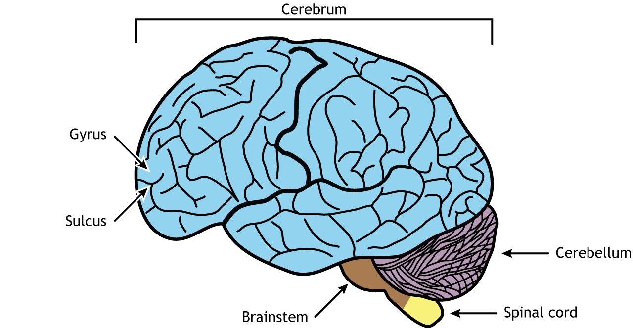
The brain is comprised of the cerebrum, cerebellum, and brainstem. The cerebrum is the most prominent region of the brain. It is divided into left and right hemispheres. The hemispheres have many of the same functions, for example, each perceives touch on one side of the body, but some functions, like language, demonstrate laterality, meaning they are primarily controlled on one side of the brain. The cerebral hemispheres in humans have many folds to increase the surface area of the brain. The ridges are called gyri and the grooves are called sulci. Large sulci are often called fissures.
The four lobes of the brain are each responsible for specific functions
Frontal Lobe
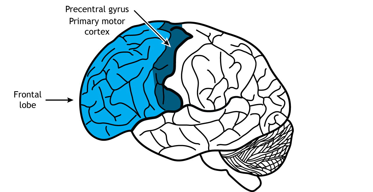
The cerebral hemispheres of the brain are divided into four lobes. The frontal lobes are the most rostral, located in the front of the brain and are responsible for higher level executive functions, like attention, critical thinking, and impulse control. They are the last brain region to fully develop, not completing development until individuals reach their 20s. The frontal lobes are also the location of the primary motor cortex, the region of the brain responsible for planning and executing movement. The primary motor cortex is located in the precentral gyrus.
View the frontal lobe using the BrainFacts.org 3D Brain
Parietal Lobe
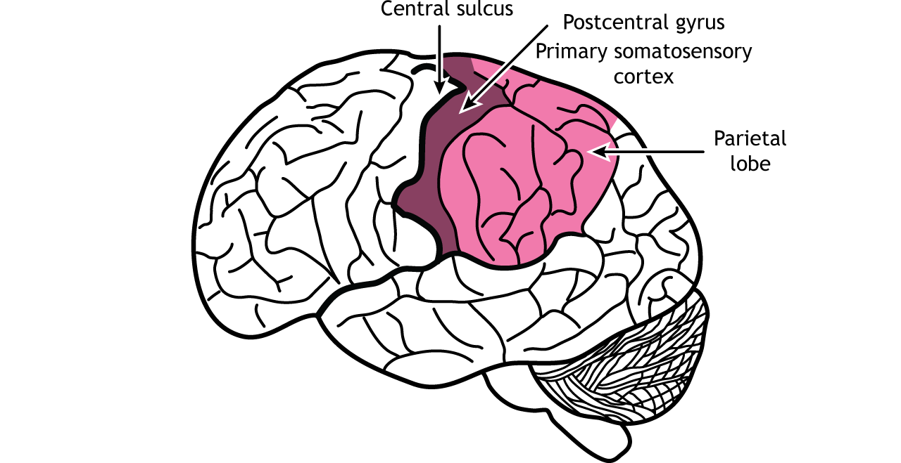
The central sulcus lies caudal to the frontal lobe and divides the frontal lobes from the parietal lobes. The parietal lobes are important for processing sensory information. The primary somatosensory cortex is located in the postcentral gyrus of the parietal lobe and is responsible for the perception of touch and pain. The parietal lobes also perform higher-level visual processing.
View the parietal lobe using the BrainFacts.org 3D Brain
Temporal Lobe
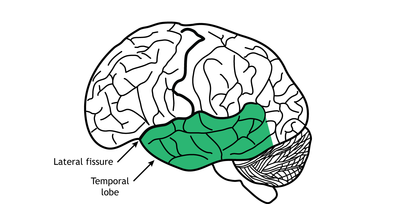
The temporal lobes are located on the side of the brain, separated from the frontal and parietal lobes by the lateral fissure. Like the parietal lobe, the temporal lobe plays a role in sensory processing, specifically with hearing, smell, taste, and higher-level visual processing. The temporal lobe is also important for speech and memory. Beneath the cerebral cortex, deep in the temporal lobes, lie the hippocampus and amygdala, two regions of the limbic system, a circuit important for emotion and memory.
View the temporal lobe using the BrainFacts.org 3D Brain
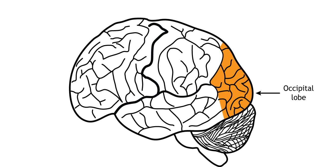
Occipital Lobe
The last lobes are the occipital lobes, the most caudal lobes located in the back of the brain. The occipital lobes’ primary function is processing of visual information.
View the occipital lobe using the BrainFacts.org 3D Brain
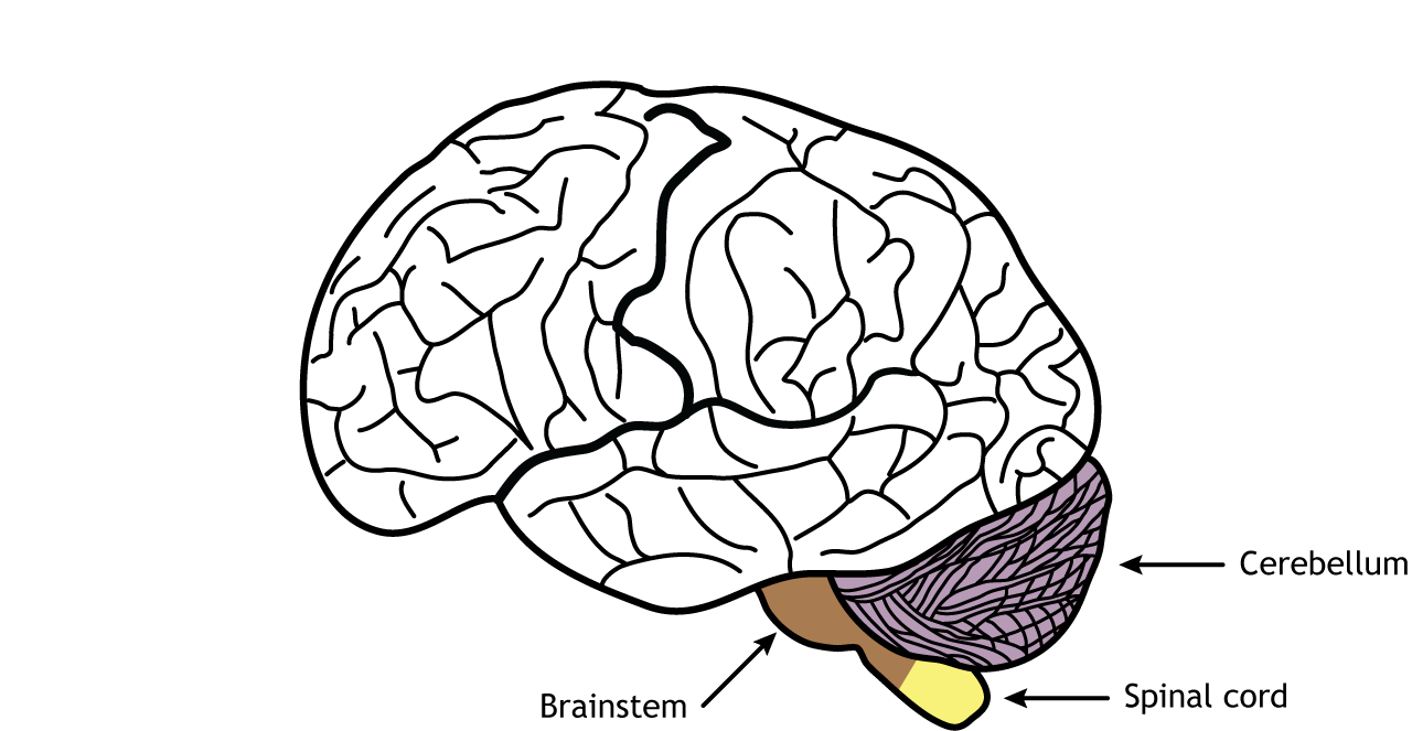
Non-Cerebral Components
The cerebellum lies inferior to the occipital lobes. The cerebellum is also divided into two hemispheres, like the cerebral cortex. The cerebellum is best known for its role in regulation and control of movement, but it is also involved in cognitive functions like emotions.
The brainstem is located between the cerebrum and the spinal cord. It is important for regulating critical functions like heart rate, breathing, and sleep. It is also the location of most of the cranial nerves.
The spinal cord, which is part of the central nervous system but not part of the brain, is responsible for receiving sensory information from the body and sending motor information to the body. Involuntary motor reflexes are also a function of the spinal cord, indicating that the spinal cord can process information independently from the brain.
View the brainstem using the BrainFacts.org 3D Brain
View the cerebellum using the BrainFacts.org 3D Brain
Landmarks on the brain can be seen from different planes of view
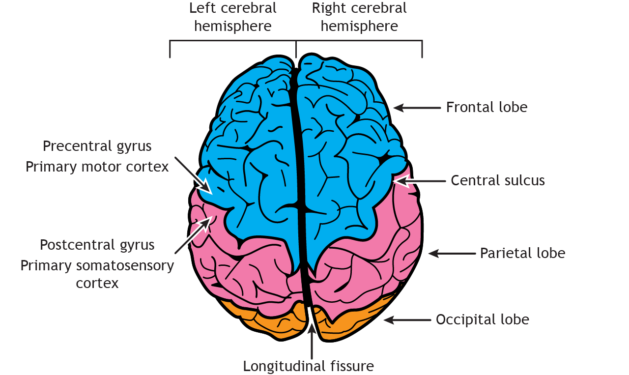
Dorsal View
Viewing the brain from above shows the bilateral symmetry of the left and right cerebral hemispheres, which are separated by the longitudinal fissure. The frontal, parietal, and occipital lobes can be seen. Similar to the lateral view, the central sulcus divides the frontal lobe from the parietal lobe. The precentral gyrus, which is the location of the primary motor cortex, sits rostral to the central sulcus, whereas the postcentral gyrus, which is the location of the primary somatosensory cortex, lies caudal to the central sulcus.
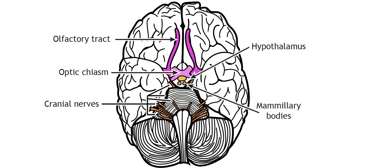
Ventral View
Underneath the brain, the frontal and temporal lobes are visible, as is the cerebellum. Like the dorsal view, the longitudinal fissure divides the cerebrum into right and left hemispheres. The pons and medulla, components of the brain stem, connect the cerebrum to the spinal cord.
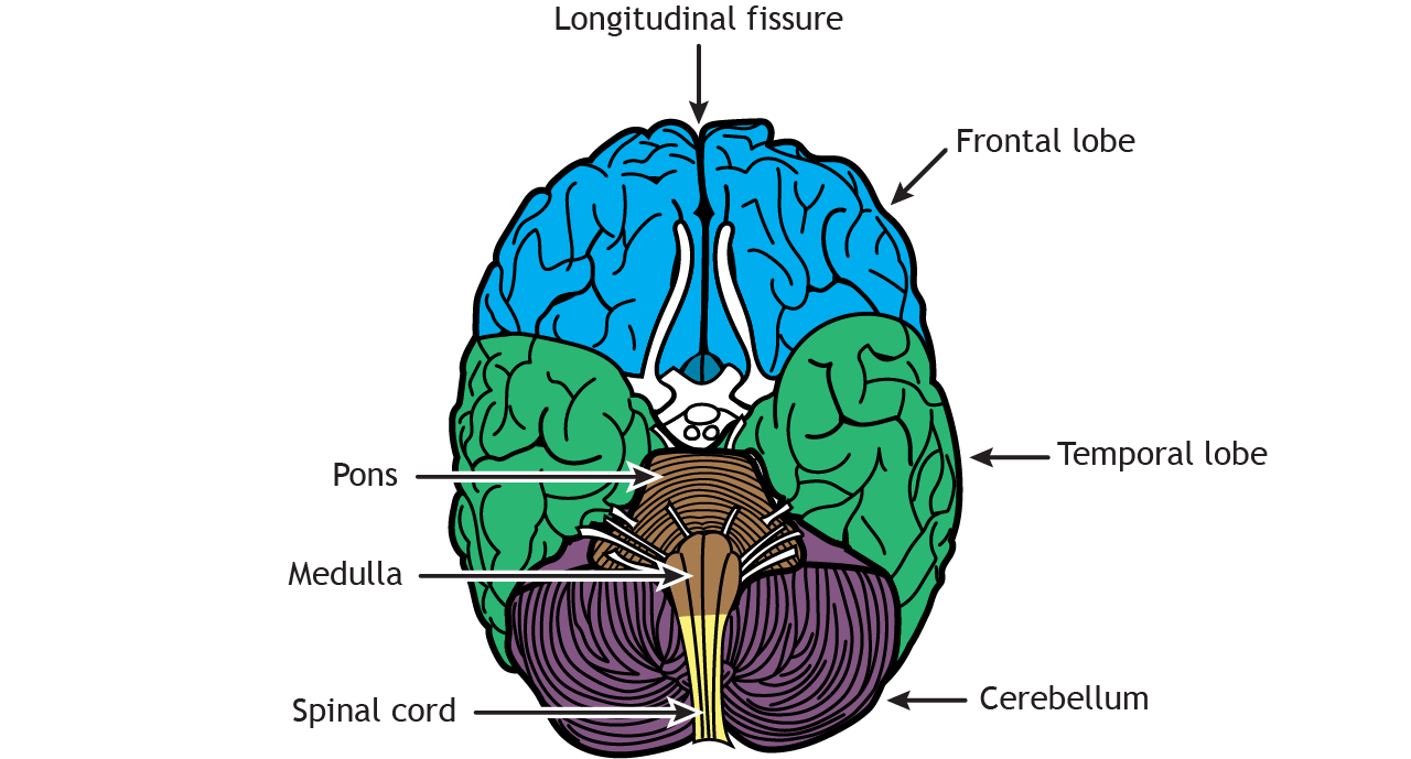
Cranial nerves are also visible on the ventral surface of the brain. The olfactory tract leads out to the olfactory bulb, which connects to the olfactory nerve. The optic tract crosses the midline at the optic chiasm, and then the optic nerve projects to the retina. Other cranial nerves enter or leave the brain at the level of the brainstem. The hypothalamus is located rostral to the pons, and the mammillary bodies project out from the hypothalamus.
Key Takeaways
- The four lobes of the cerebral cortex each have specific functions
- The cerebral cortex has gyri and sulci to increase the surface area
- The cerebral cortex, underlying structures, cerebellum, brainstem, and spinal cord form the central nervous system
Test Yourself!
