
22 Vision: Central Processing
Visual Fields
Before learning the pathway that visual information takes from the retina to the cortex, it is necessary to understand how the retina views the world around us. The full visual field includes everything we can see without moving our head or eyes.
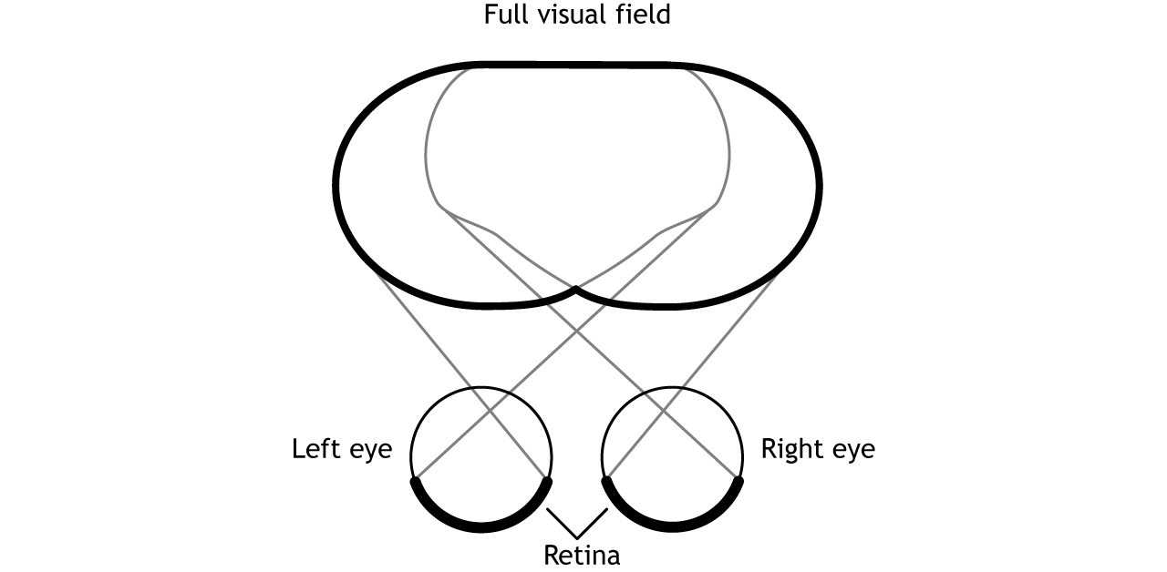
Visual fields can be divided in multiple ways, each dependent on different regions of the retina
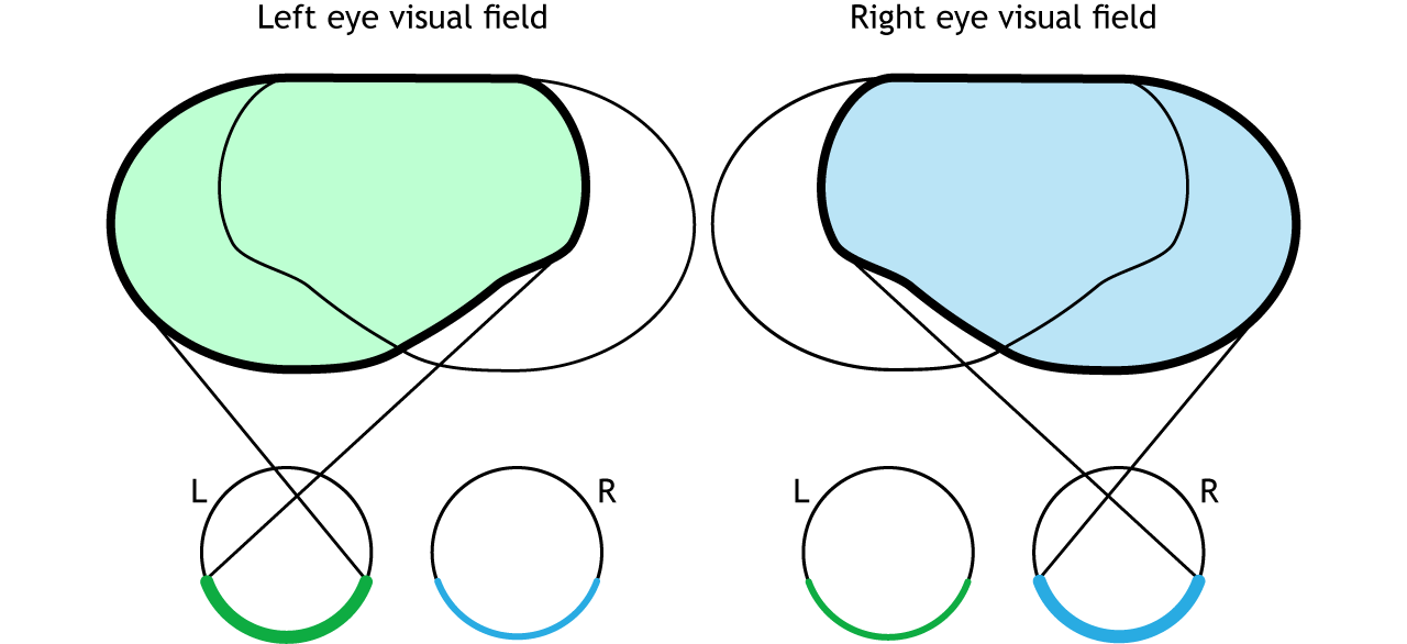
The full visual field can be divided in a few ways. Each individual eye is capable of seeing a portion of, but not the entire, visual field.
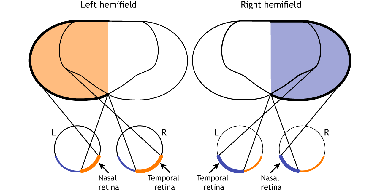
The full visual field can also be divided into the right and left hemifields. The hemifields range from the most peripheral point to the center point, splitting the full visual field into two equal regions. Both eyes are involved in viewing each hemifield. The fovea separates the retina into two sections: the nasal retina and the temporal retina. The nasal retina is the medial portion that is located toward the nose. The temporal retina is the lateral portion that is located toward the temples and temporal lobe. The nasal retina from one eye along with the temporal retina from the other eye are able to view an entire hemifield.
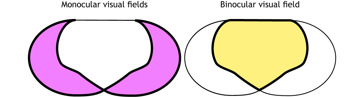
Finally, the full visual field can be separated into monocular and binocular regions. Each monocular field is visual space that can only be viewed by one eye. The binocular region is visual space that can be viewed by both eyes.
Pathway to Brain
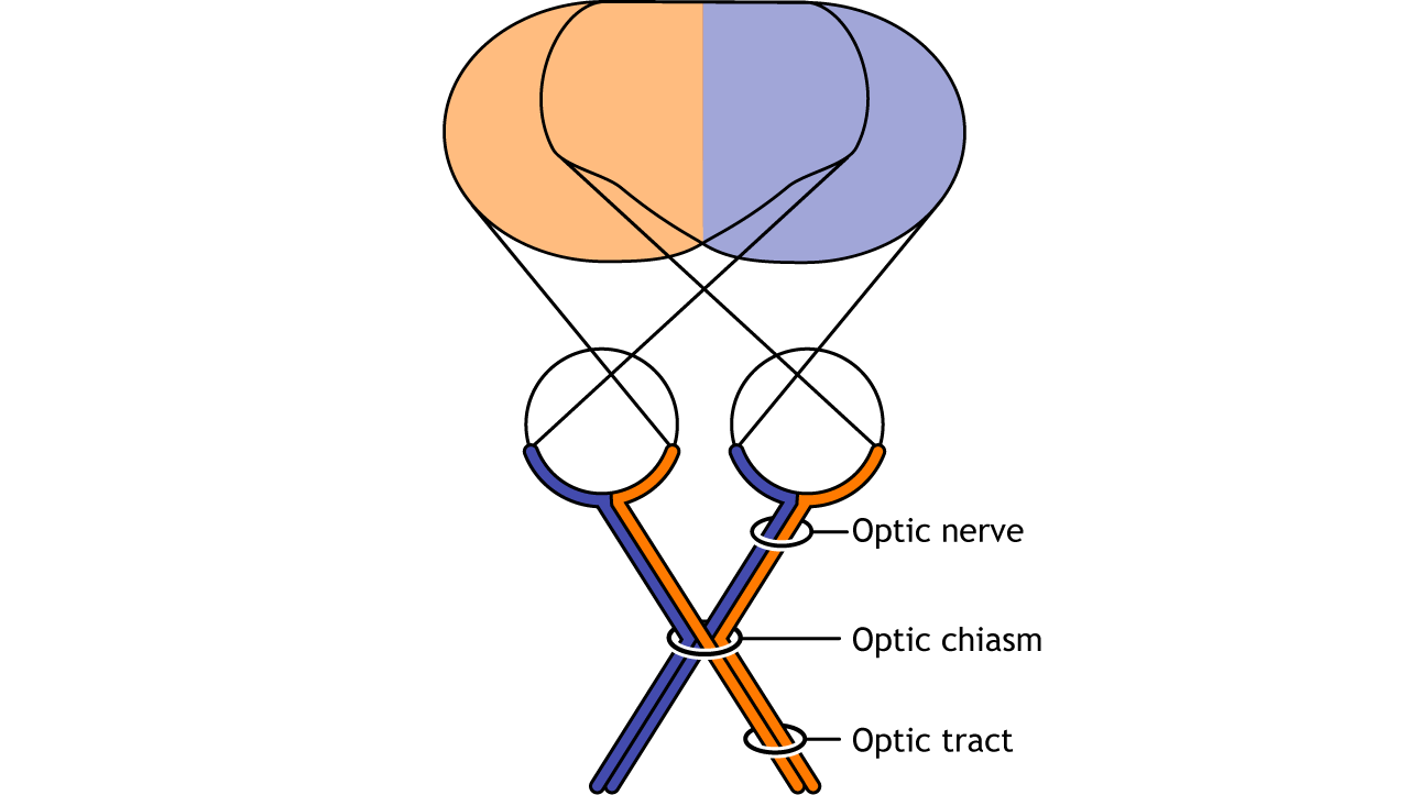
The right visual field is processed by the left side of the brain; the left visual field is processed by the right side of the brain
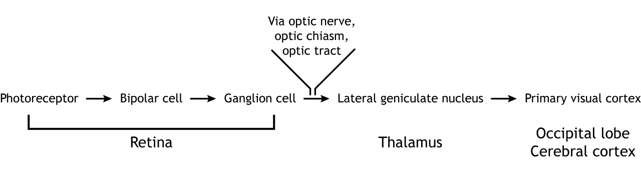
Visual information from each eye leaves the retina via the ganglion cell axons at the optic disc, creating the optic nerve. Prior to entering the brain, axons from the nasal portion of each retina cross the midline at the optic chiasm. Since the axons from the nasal retina cross to the opposite side of the nervous system but the temporal retina axons do not, this leads to the brain processing input from the contralateral (opposite side) visual hemifield. Therefore, the right side of the brain receives visual information from the left hemifield and vice versa.
View the optic nerve (cranial nerve II) using the BrainFacts.org 3D Brain
The optic tract enters the brain and ascends to synapse in the lateral geniculate nucleus of the thalamus. From there, axons project to the primary visual cortex, also called the striate cortex or V1, located in the occipital lobe.

View the thalamus using the BrainFacts.org 3D Brain
View the primary visual cortex using the BrainFacts.org 3D Brain
Receptive Fields
Receptive fields transition to circular to lines and then to more complex forms (like faces), orientation, and direction of movement
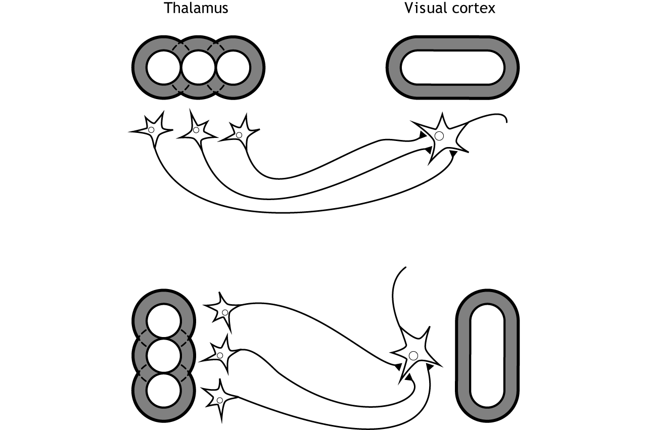
As information moves from the retina to the cortex, receptive fields become larger and more complex. Receptive fields in the thalamus continue to be circular in shape like the receptive fields of the retinal neurons. However, once information reaches the primary visual cortex, these circular receptive fields combine to create receptive fields that are activated by lines.
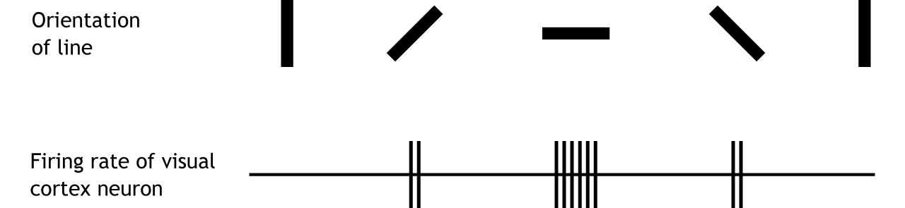
These receptive fields cause neurons in the primary visual cortex to respond best to a line in a specific orientation. The firing rate of the neuron will increase as the line rotates toward the “preferred” orientation. The firing rate will be highest when the line is in the exact preferred orientation. Different orientations are preferred by different neurons.
Higher-Level Processing of Sensory Information
Sensory system processing of input does not end upon reaching the primary sensory cortex in any sensory system. Information typically gets sent from the primary sensory cortex to other sensory association regions throughout the brain. The characteristics of sensory information becomes more complex as this higher-level processing occurs.
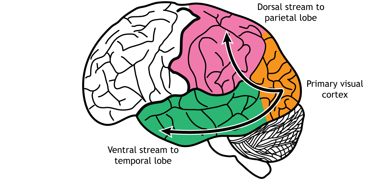
Beyond the Primary Visual Cortex
The dorsal stream recognizes movement; the ventral stream recognizes objects
In the visual system, there are two broad streams of information that leave the primary visual cortex. Information that travels from the primary visual cortex down through the inferior temporal lobe is responsible for determining object recognition, or what an object is. Differentiating between an apple and a person occurs in this stream. Information that travels from the striate cortex up through the parietal lobe is responsible for motion or spatial components of vision.
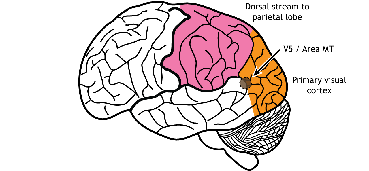
Dorsal Stream – “Where is it?”
One of the most important regions in the dorsal pathway is region MT, also called V5. In this region, neurons are preferentially activated by a specific direction of movement by an object – for example, left to right or up to down. As an example, remember the receptive fields in the primary visual cortex were activated by lines at a specific orientation. Like that, in V5, the neurons would be activated by lines moving in a specific direction.
As information continues to be processed through the dorsal stream, the neurons become selective for more complex motions. The dorsal stream is also important for processing our actions in response to visual stimulation, for example, reaching for an object in the visual field or navigating around objects while walking.
Ventral Stream – “What is it?”
Object identification is a key function of our visual system. The ventral visual stream is responsible for this process. Like the more complex activation characteristics of region MT in the dorsal stream, neurons in Area V4 in the ventral stream show more complex receptive fields and show sensitivity to shape and color identification. As visual information continues to be processed through the inferior temporal lobe, differentiation of objects occurs. For example, in a region called the fusiform face area, located in the fusiform gyrus, which lies on the ventral aspect of the temporal lobe, neurons are activated by faces and can be specialized to categories of faces, like those people familiar to you versus strangers.

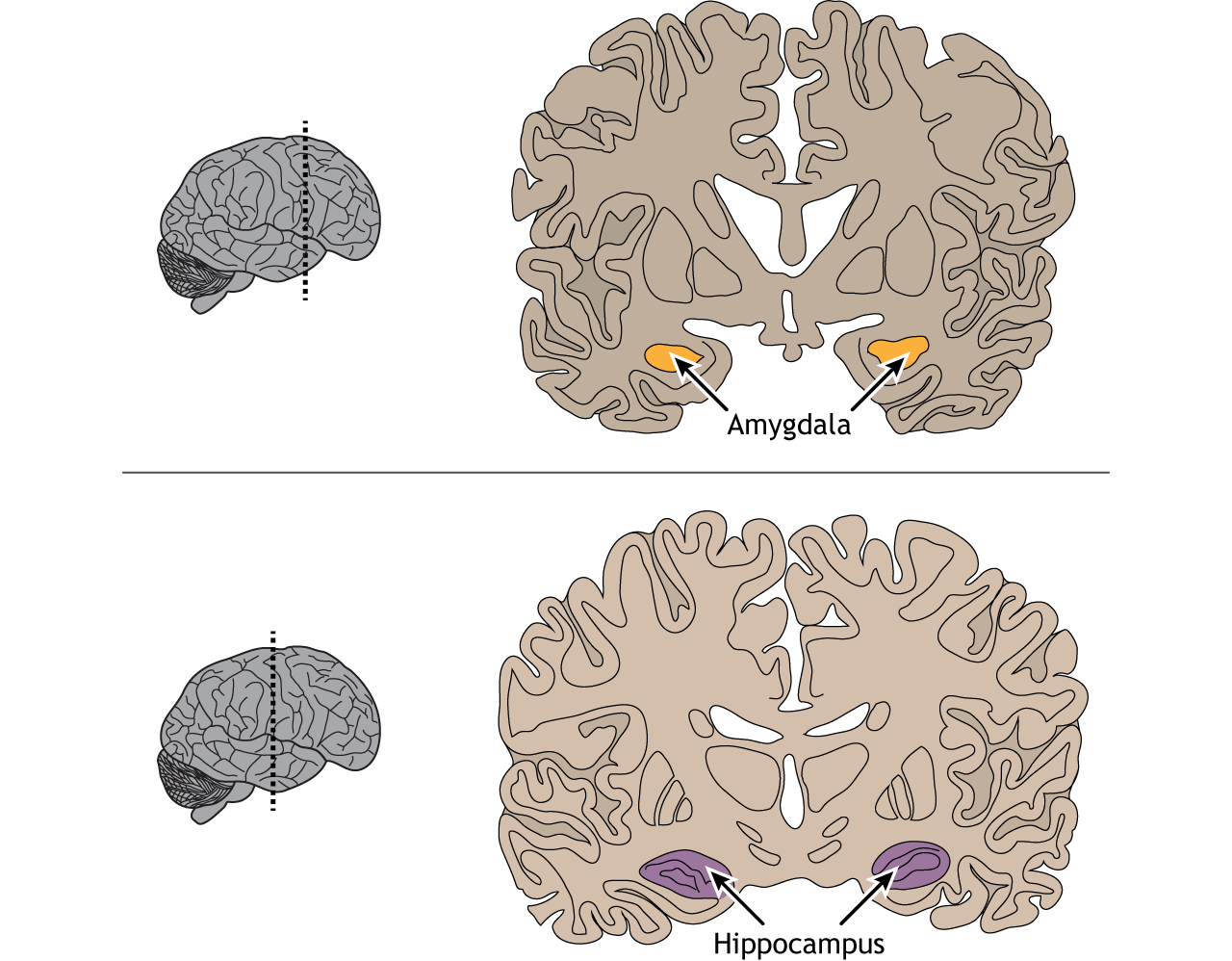
The inferior temporal lobe also makes reciprocal connections with the structures in the limbic system. The limbic system plays an important role in processing emotions and memory, both of which are significant components to visual perception. The amygdala ties visual stimuli with emotions and provides value to objects. A family member will have emotional ties that a stranger will not. The hippocampus is responsible for learning and memory and helps establish memories of visual stimuli.
View the amygdala using the BrainFacts.org 3D Brain
View the hippocampus using the BrainFacts.org 3D Brain
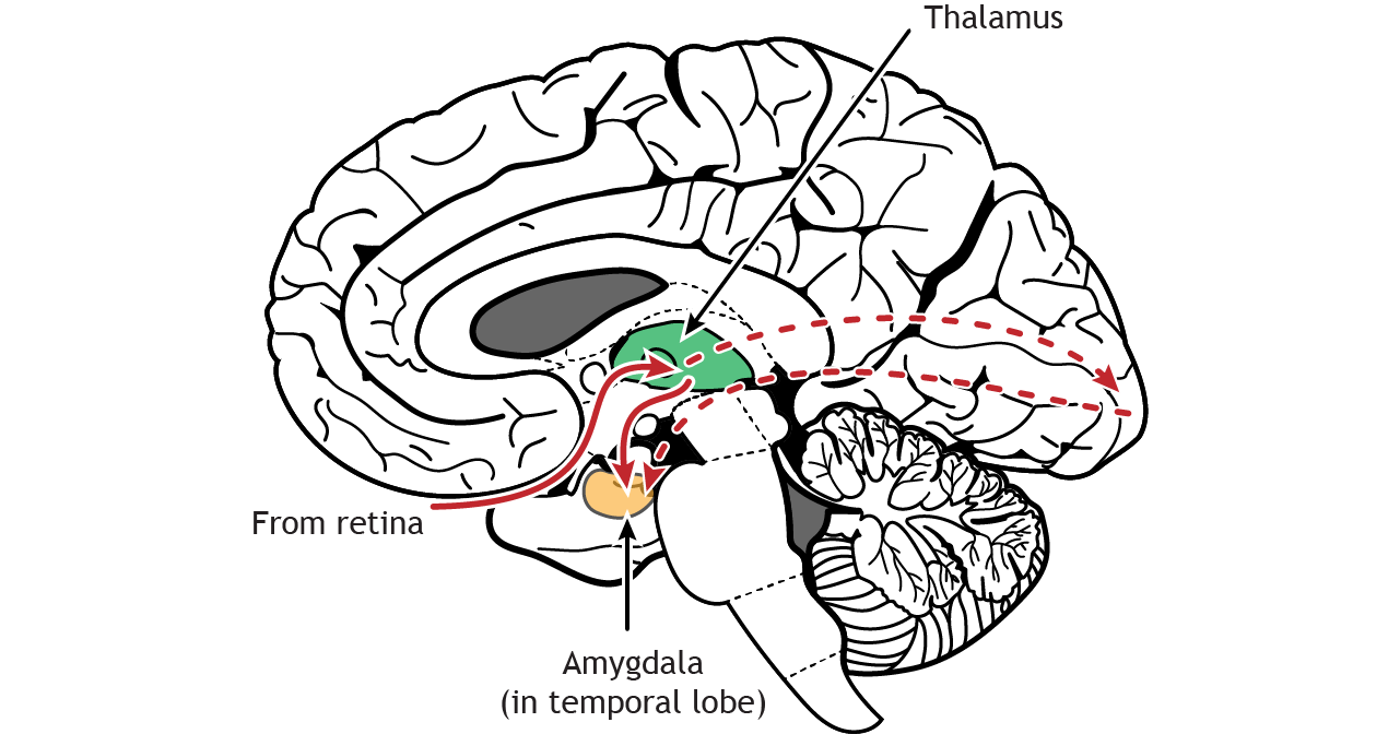
Pathways through the Amygdala
The amygdala receives visual information through multiple pathways
In addition to the projections to the amygdala via the ventral stream, the amygdala also appears to rely on input from the thalamus that is independent of the ventral stream pathway. A shorter pathway travels from the retina to the amygdala via the thalamus. It is believed that this pathway allows for a rapid responses to the threats and allows visual stimuli to activate the amygdala quicker than processing through the visual cortex. In fact, studies have shown that pictures of angry or fearful faces can cause amygdala activation without conscious awareness of seeing the image, a result of images being shown for only milliseconds to the subject.
Non-Thalamic Pathways
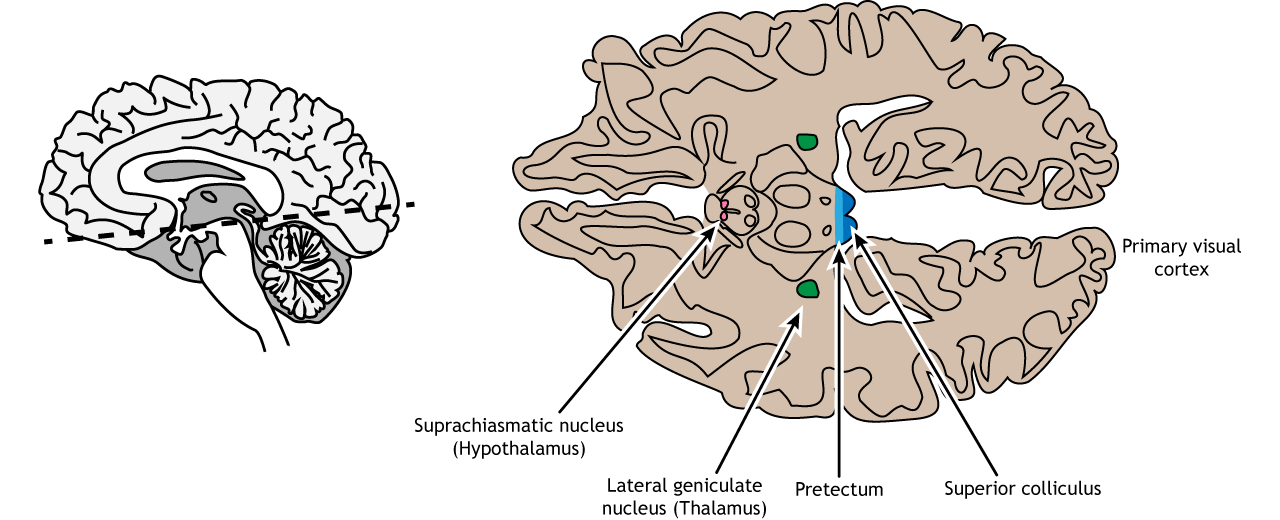
The retina sends projections to more regions than just the thalamus
Although most retinal output projects to the lateral geniculate nucleus of the thalamus and then to the primary visual cortex, there are some axons that project to other areas of the brain. A subset of specialized retinal ganglion cells project to the suprachiasmatic nucleus in the hypothalamus. This region is critical for circadian rhythms and the sleep/wake cycle. Other retinal neurons send axons to the pretectum, a midbrain region that communicates with motor nuclei and is responsible for pupillary control. Finally, other ganglion cells project to the superior colliculus, another midbrain region. This pathway is responsible for movements that will orient the head and eyes toward an object to focus the object in the center of the visual field, the region of highest visual acuity.
View the hypothalamus using the BrainFacts.org 3D Brain
View the midbrain using the BrainFacts.org 3D Brain
Key Takeaways
- The nasal and temporal retinal regions are responsible for viewing specific regions of the visual field
- Some retinal projections cross the midline at the optic chiasm, causing the left side of the brain to process the right visual hemifield and vice versa
- The retinal axons synapse in the lateral geniculate nucleus of the thalamus. Information then travels to the primary visual cortex
- Receptive fields and the preferred visual stimuli for neuron activation become more complex as information moves through the visual pathway
- Retinal cells and thalamic neurons have circular receptive fields with inhibitory surround
- Primary visual cortex neurons have linear receptive fields are are activated by a line in a specific orientation
- Area MT / V5 is activated by motion in a specific direction
- Area V4 is activated by specific shapes and colors
- The fusiform gyrus is activated by faces
- The retina also projects to midbrain regions
Test Yourself!
