
27 Taste
Being able to sense chemicals in the environment through taste and olfaction can help an organism find food, avoid poisons, and attract mates. Humans can perceive five basic tastes: salty, sour, bitter, sweet, and umami. Bitter taste often indicates a dangerous substance like a poison, sweet taste signifies a high energy food, salty taste indicates a substance with high salt content, sour taste indicates an acidic food, and umami taste indicates a high protein food.
Tongue anatomy
The surface of the tongue is covered in small, visible bumps called papillae. Taste buds are located within the papillae, and each taste bud is made up of taste receptor cells, along with supporting cells and basal cells, which will eventually turn into taste receptors cells. The taste cells have a lifespan of approximately two weeks, and the basal cells replace dying taste cells. The taste cells have microvilli that open into the taste pore where chemicals from the food can interact with receptors on the taste cells. Although taste cells are not technically neurons, they synapse and release neurotransmitters on afferent axons that send taste perception information to the brain.
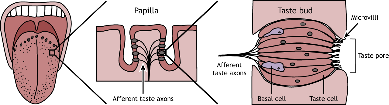
The entire tongue is capable of perceiving all five tastes, meaning there are taste receptors for each taste present across the entire surface. However, some regions of the tongue have a slightly lower threshold to respond to some tastes over others. The tip of the tongue is the region most sensitive to sweet, salt, and umami tastes. The sides are most sensitive to sour, and the back of the tongue to bitter tastes.

Taste transduction
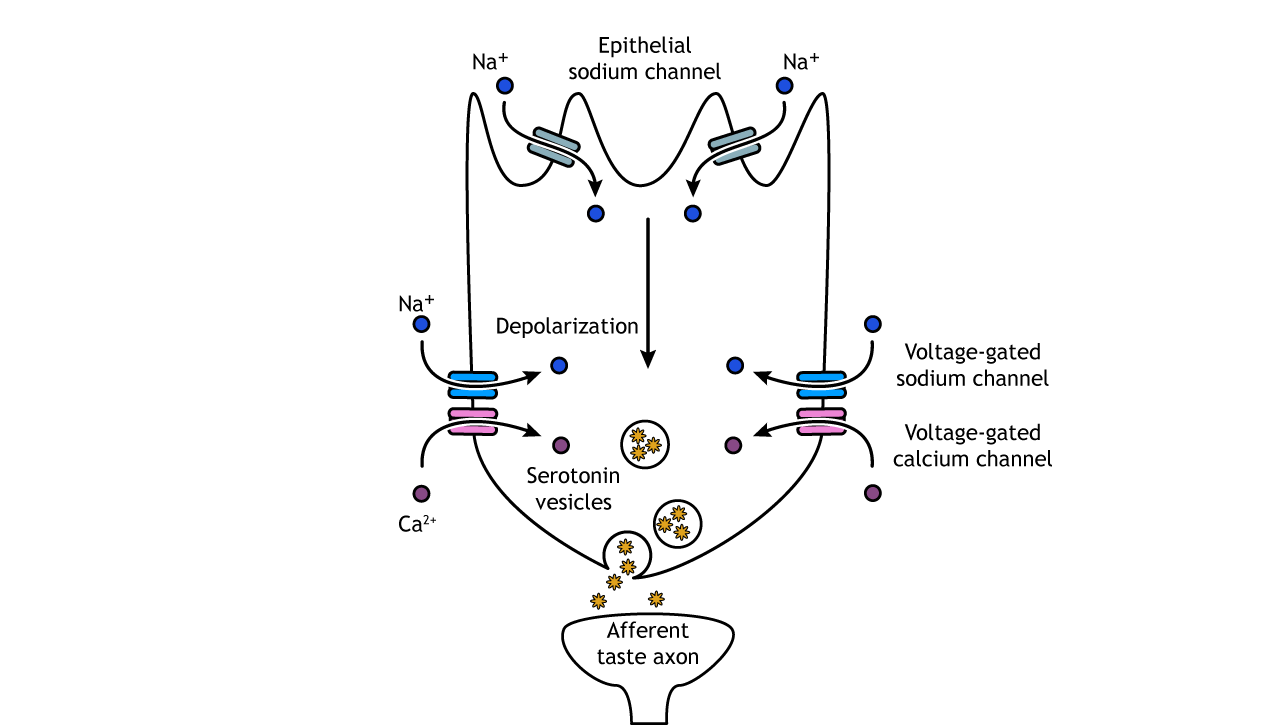
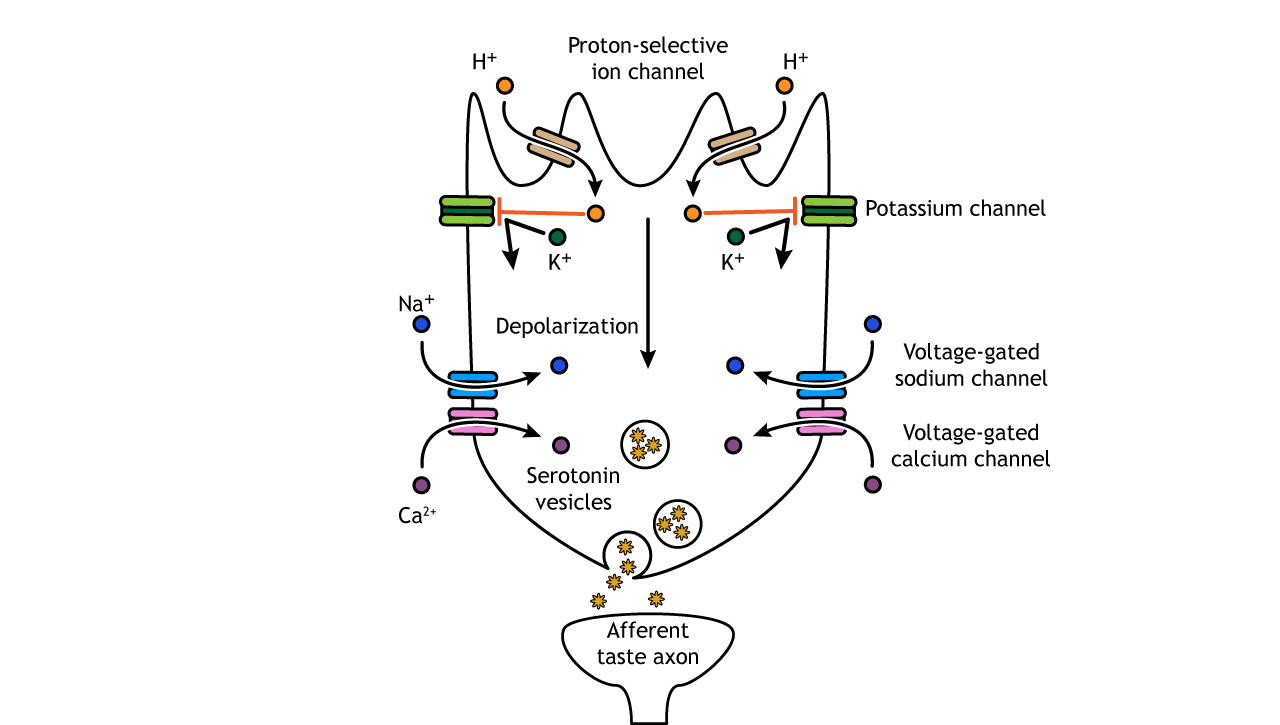
Salt and sour taste receptor cells depolarize through the movement of ions into the cell body and then release serotonin
Salt
Salt taste is mediated by the presence of epithelial sodium channels. These receptors are usually open, and when foods are ingested with high salt concentrations, sodium flows into the cell causing a depolarization. This change in membrane potential opens voltage-gated sodium and calcium channels. The increased calcium influx causes the release of serotonin-filled vesicles. The serotonin acts on the afferent taste axon causing depolarization and action potentials.
Sour
Foods taste sour because of their acidity, and when acids are present in water, they produce hydrogen ions (protons). The exact mechanism for sour taste transduction has yet to be worked out, but it is believed that protons enter the cell through an ion channel, and then block potassium channels. The decreased efflux of potassium, along with the presence of the protons, depolarizes the cell causing voltage-gated sodium and calcium channels to open. Like salt taste transduction, the increase in intracellular calcium causes release of serotonin into the synapse.
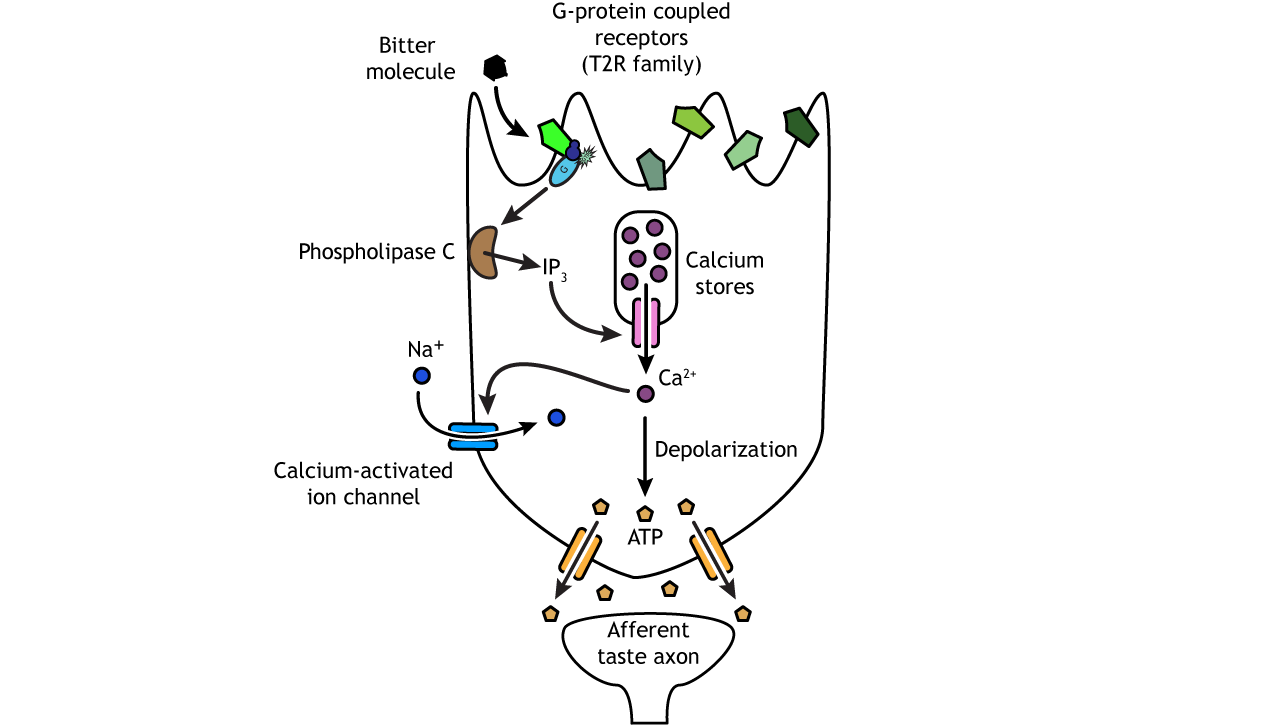
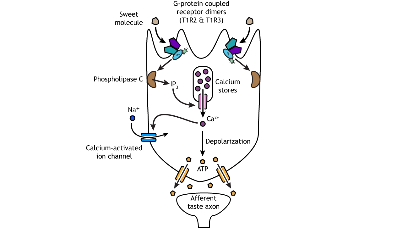
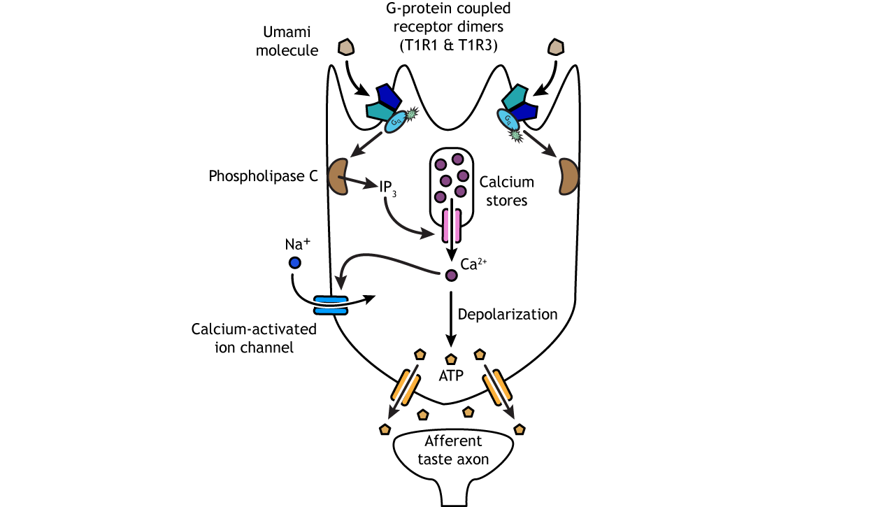
Bitter, sweet, and umami taste receptor cells depolarize through activation of G-protein coupled receptors and then release ATP
Bitter
Bitter, sweet, and umami compounds all activate taste receptor cells via G-protein coupled receptors. The bitter receptors are from the T2R family of receptor proteins; humans have over 25. Each taste cell can express most or all of the different receptor types, allowing for the detection of numerous molecules, which is important when wanting to avoid dangerous substances like poisons and toxins.
Activation of the G-protein receptor uses a second messenger system to increase intracellular calcium, which opens ion channels, allowing the influx of sodium. These ion changes depolarize the cell and cause ATP-specific channels to open, allowing ATP to enter the synapse and act on the afferent taste axon.
Sweet
Sweet and umami receptors are comprised of G-protein coupled dimers, meaning two separate proteins function together as one. The receptors are encoded by the T1R family of receptor proteins. Sweet receptors are dimers of the T1R2 and T1R3 proteins. Both proteins need to be present and functioning for activation of a sweet taste cell. Like bitter cells, activation of the G-protein receptor uses a second messenger system to release calcium from intracellular stores and increase the influx of sodium. These ion changes depolarize the cell and cause ATP-specific channels to open, allowing ATP to enter the synapse and act on the afferent taste axon.
Umami
Umami receptors are comprised of the T1R3 protein, like the sweet receptor, but it is paired with the T1R1 protein. Once the G-protein coupled receptor is activated, the transduction pathway is the same as bitter and sweet taste cells.
Coding Properties of Taste
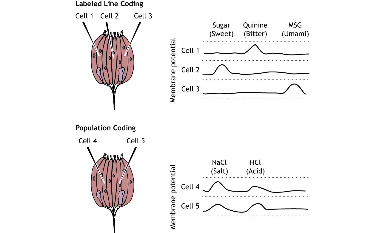
At the level of the taste receptor, the type of taste perceived depends primarily on which receptor type is activated.
Of the five tastes, only two neurotransmitters are used to communicate information to the central nervous system, so how does our brain know what tastes to perceive? The answer is how the information is encoded. Most taste cells use a labeled line coding method, which means that each cell and the related afferent taste axon only responds to one type of taste. For example, bitter cells only express bitter receptors and are only activated by bitter molecules. These bitter taste cells activate bitter sensory neurons and bitter regions of the taste cortex. A small portion of taste cells do use population coding as well, meaning more than one tastant can activate the cell, and perception is based on a combination of multiple cells each with a different response. Most information, however, is encoded via labeled line at the level of the taste cell. As taste is processed through higher levels of the brain, however, population coding becomes more important in the perception of taste.
Mouth and Throat Anatomy
Although taste receptor cells are most prevalent on the tongue, there are other regions of the mouth and throat, including the palate, pharynx, and epiglottis, that also are sensitive to food and play a role in taste perception. The olfactory system is tightly linked to our sense of taste as well, and odorant compounds from food can reach odor receptors in the nasal cavity.

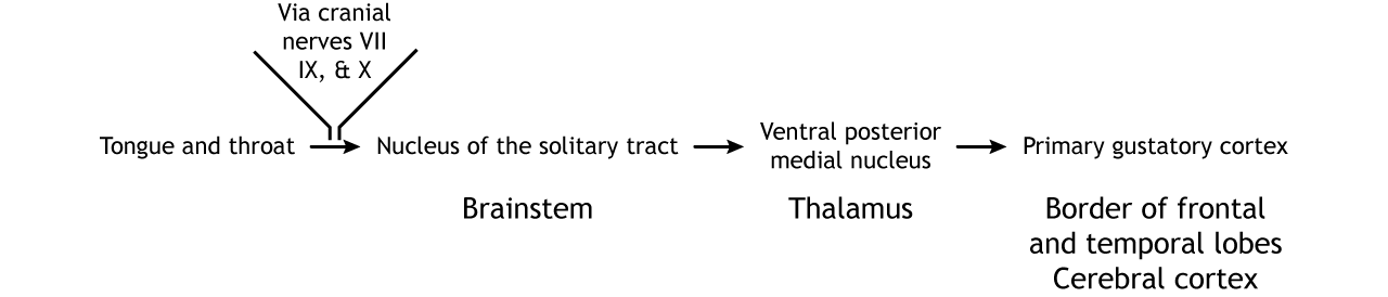
Pathway
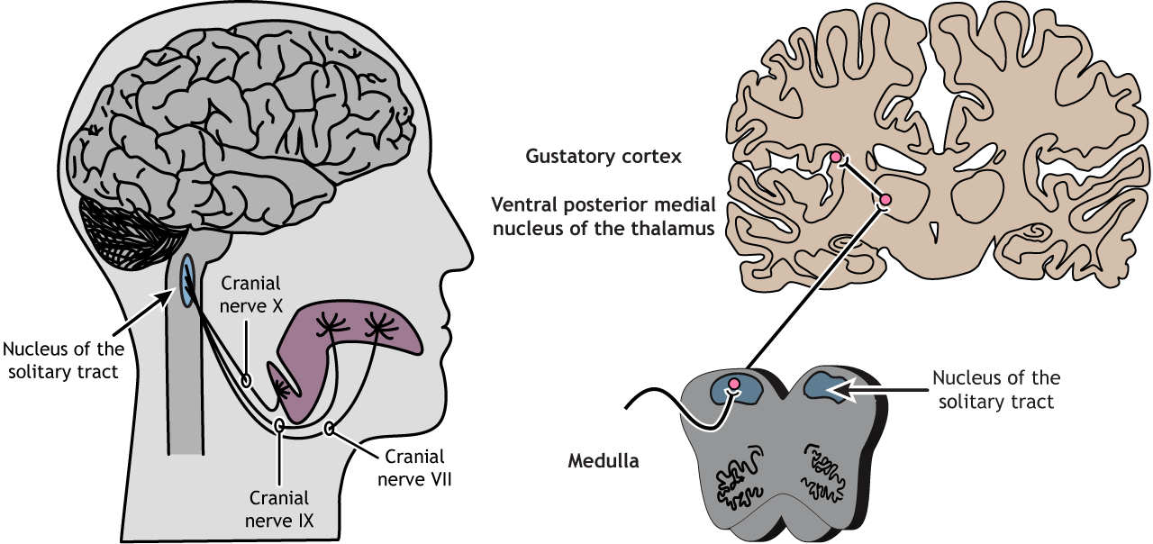
Cranial nerves VII, IX, and X process taste information
The tongue is innervated by three cranial nerves. The front two-thirds of the tongue is innervated by cranial nerve VII. The back third is innervated by cranial nerve IX. Finally, the epiglottis and pharynx are innervated by cranial nerve X. All three cranial nerves enter the brainstem at the medulla and synapse in the nucleus of the solitary tract. From there, information is sent to the ventral posterior medial nucleus of the thalamus. Thalamic neurons send projections to the gustatory cortex. The gustatory cortex is located deep in the lateral fissure in a region called the insula. Information processing taste stays primarily on the ipsilateral side of the nervous system. Projections within the brain also exist between the taste regions and the hypothalamus and amygdala.
View the facial nerve (cranial nerve VII) using the BrainFacts.org 3D Brain
View the glossopharyngeal nerve (cranial nerve IX) using the BrainFacts.org 3D Brain
View the vagus nerve (cranial nerve X) using the BrainFacts.org 3D Brain
View the thalamus using the BrainFacts.org 3D Brain
Flavor
How do 5 basic tastes turn into the myriad complex taste sensations we experience when eating food? Olfaction plays an important role in the perception of flavor, as do vision and touch. Taste information combines with information from these other sensory systems in the orbitofrontal cortex located in the frontal lobe. This region is believed to be important for the pleasant and rewarding aspects of food. Additionally, as taste is processed in higher-order regions of the CNS, information is combined using population coding mechanisms. Test how your senses combine to create flavor at home!
View the orbitofrontal cortex using the BrainFacts.org 3D Brain
Key Takeaways
- Taste cells express specific taste receptors and are located in taste buds within the papillae
- Salt and sour taste cells rely on ion channels to depolarize the cell and release serotonin
- Bitter, sweet, and umami taste cells rely on G-protein coupled receptors and second messengers that open ATP channels
- At the level of the taste receptor cells, taste is perceived by using labeled line coding
- Multiple regions in the mouth and throat play a role in processing of taste
- Three cranial nerves innervate the tongue and throat
- The cranial nerves synapse in the nucleus of the solitary tract in the medulla. Information then travels to the ventral posterior medial nucleus of the thalamus and then to the gustatory cortex
- To perceive complex flavors, information from other sensory systems is combined with taste information in the orbitofrontal cortex
Test Yourself!
