
19 Brainstem and Spinal Cord
Brainstem
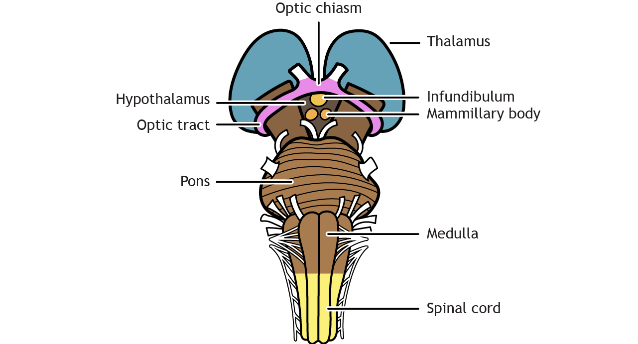
The brainstem is made up of the midbrain, pons, and medulla. It is located between the diencephalon (thalamus and hypothalamus region) and the spinal cord. All connections between the brain and the body must travel through the brainstem. The brainstem also plays an important role in the regulations of consciousness and regulates critical functions like heart rate and breathing.
The mammillary bodies are located on the ventral side of the hypothalamus. The infundibulum is the stalk between the hypothalamus and the pituitary and is located rostral to the mammillary bodies. The optic tract leaves the diencephalon and crosses the midline at the optic chiasm.
Cranial Nerves
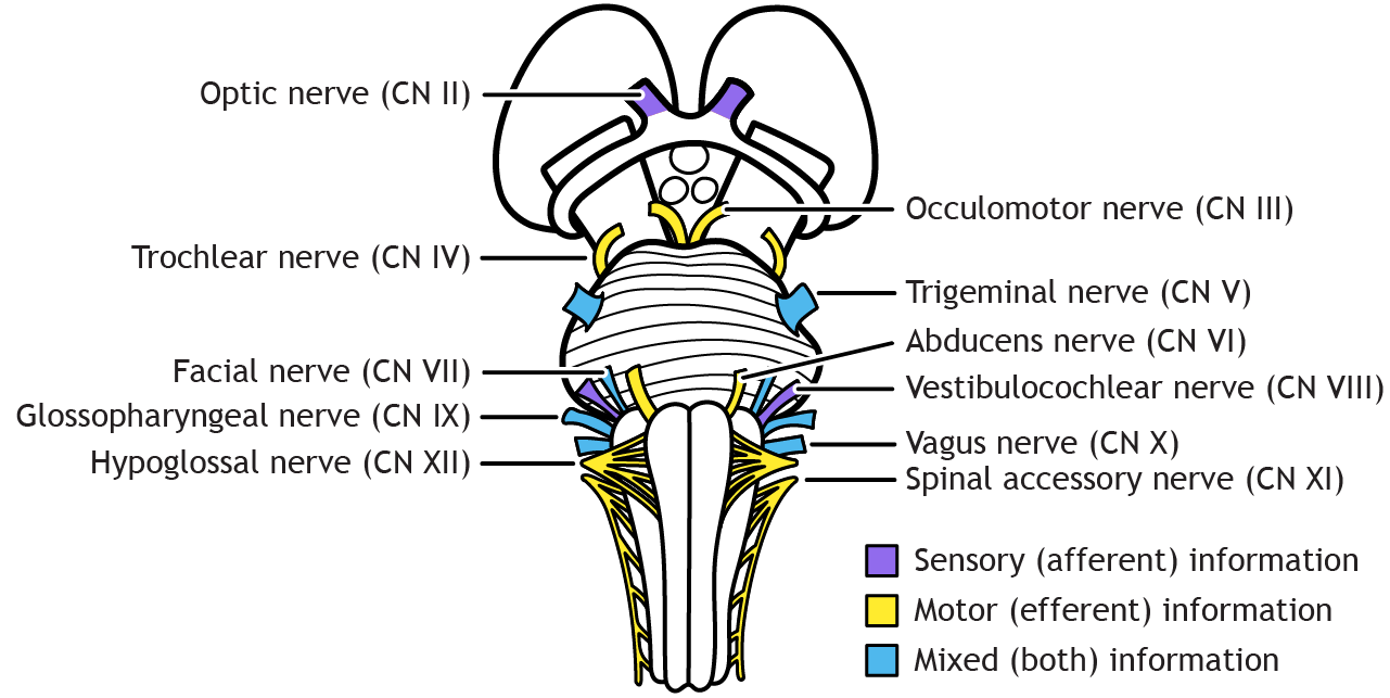
A critical component of the brainstem is the presence of the twelve pairs of cranial nerves, which provide sensory and motor innervation to the head, face, and neck, as well as autonomic innervation to the organs in the abdomen. The first two cranial nerves (olfactory [I] and optic [II]) are part of the central nervous system. They carry sensory information, smell and vision, respectively. They enter the forebrain and not the brainstem. The remaining nerves, like spinal nerves, have their axons in the peripheral nervous system. The oculomotor [III] and trochlear nerves [IV], whose functions are to move the eye, exit the brainstem from the midbrain. The trochlear nerve is the only cranial nerve to exit on the dorsal surface of the brainstem. The trigeminal nerve [V] is the largest cranial nerve and carries both sensory and motor information from the face; axons in this nerve enter and exit from the pons. The abducens nerve [VI], another eye movement nerve, exits at the junction of the pons and medulla, as do the facial [VII] and vestibulocochlear [VIII] nerves. The facial nerve contains both sensory and motor axons, whereas the vestibulocochlear nerve carries only sensory information related to hearing and balance. The glossopharyngeal [IX] and vagus [X] nerves also carry both sensory and motor information and enter/exit from the medulla. The glossopharyngeal nerve is responsible for movement of the throat muscles and taste; the vagus nerve is the primary autonomic cranial nerve and contains parasympathetic fibers that innervate the heart, lungs, and abdominal organs. Finally, the spinal accessory [XI] and hypoglossal [XII] are motor nerves with the hypoglossal exiting the brainstem from the medulla and the spinal accessory nerve exiting from the cervical spinal cord. The spinal accessory innervates muscles in the throat, shoulder, and neck, and the hypoglossal innervates muscles of the tongue.
Spinal Cord
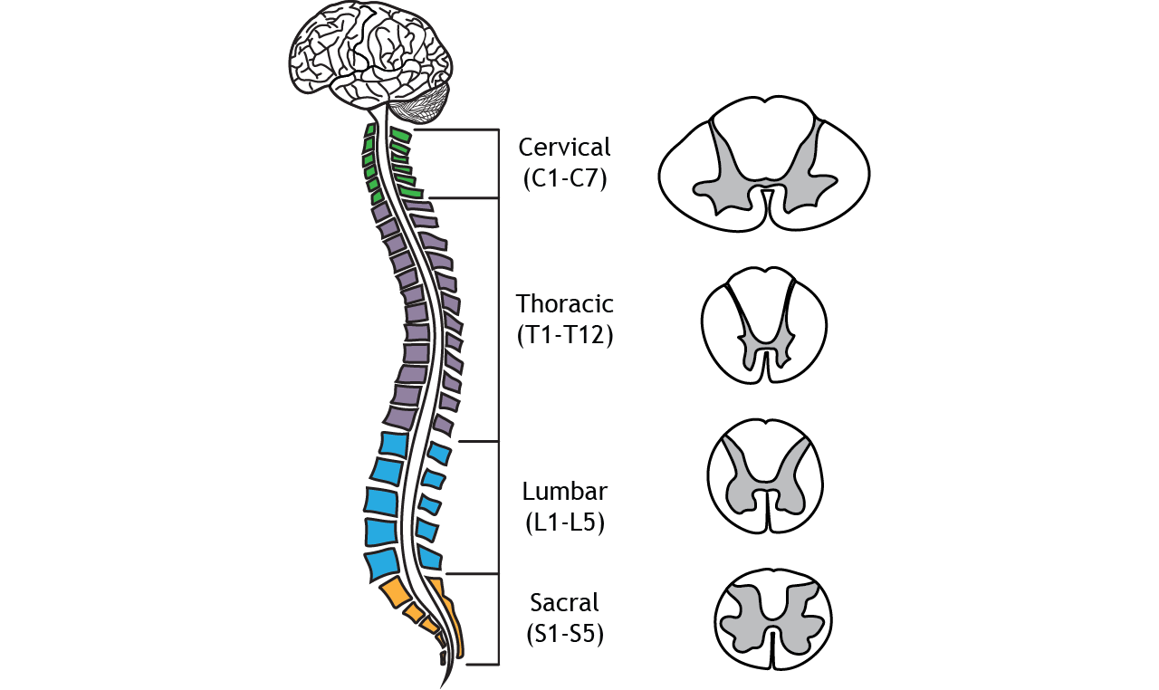
The spinal cord begins at the base of the brainstem. The vertebral column is divided into four main regions: cervical, thoracic, lumbar, and sacral. The spinal cord and spinal nerves that enter and exit the vertebral column are divided into these regions as well. The cervical division consists of seven segments (C1-C7), the thoracic consists of twelve segments (T1-T12), the lumbar consists of five segments (L1-L5), and the sacral division consists of five segments (S1-S5). The shape of the spinal cord changes over the length of the vertebral column, a result of the function of the spinal nerves. For example, motor information to the hands and arms, so the region where motor neurons are located (ventral horn) is larger than segments with minimal motor output.
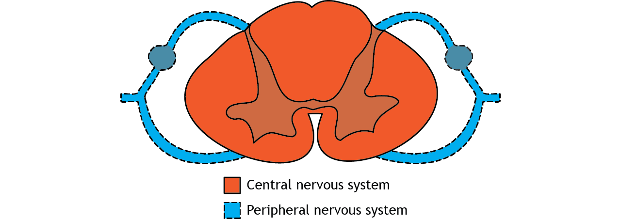
The spinal cord is part of the central nervous system, but the fibers that leave and enter the spinal cord are located in the peripheral nervous system. These spinal nerves can then extend to or from target tissue throughout the body.
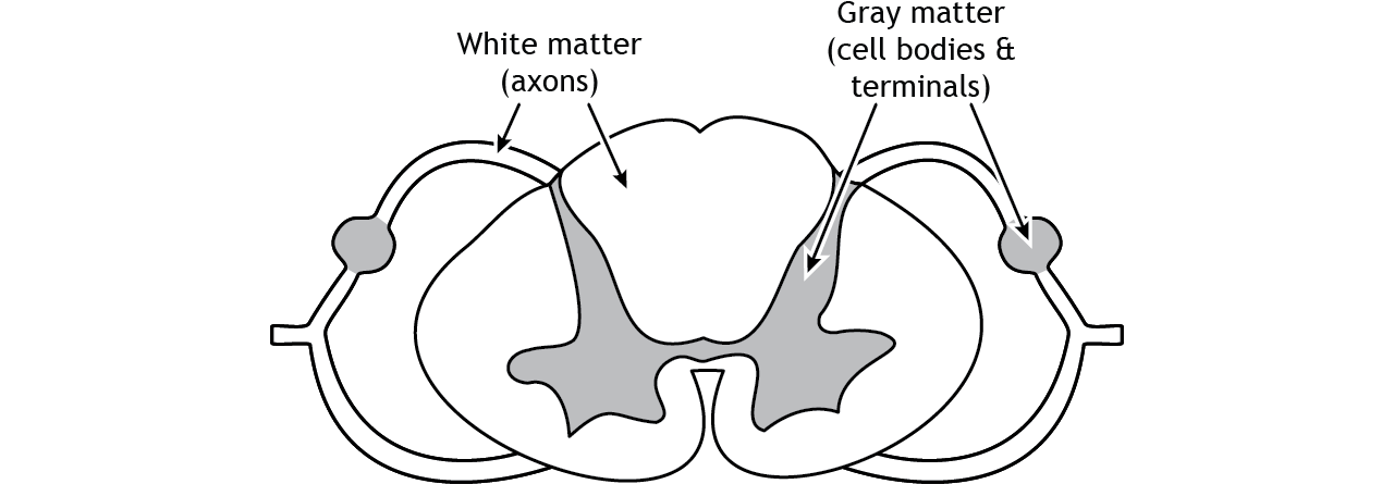
Like the brain, the spinal cord is also made up of regions of white matter and gray matter. White matter regions are comprised of axons. It appears white due to the myelin sheath on the axons. Gray matter regions are comprised of cell bodies and dendrites. Gray matter is the location of most synapses.
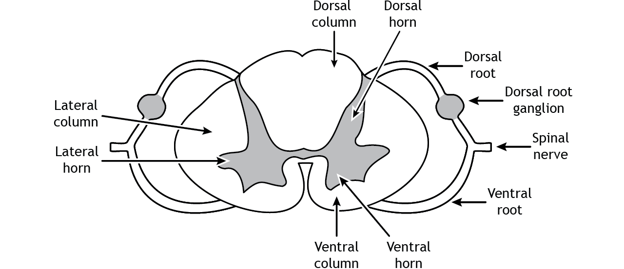
The white matter in the spinal cord is divided into structures called columns because the axons in these regions are either ascending toward the brain or descending toward the appropriate spinal nerve. The dorsal column is on the dorsal or posterior side of the spinal cord, the ventral column is on the ventral or anterior side of the spinal cord, and the lateral column lies between them. The gray matter is likewise divided into regions called horns. The dorsal horn is the location of sensory synapses, the ventral horn is the location of motor neuron cell bodies, and the lateral horn is the location of cell bodies of the autonomic nervous system. The dorsal root and ventral root consist of the axons of afferent (dorsal) and efferent (ventral) fibers. They combine to form the spinal nerves. Sensory neuron cell bodies are located in the dorsal root ganglion, a gray matter region of the dorsal root.
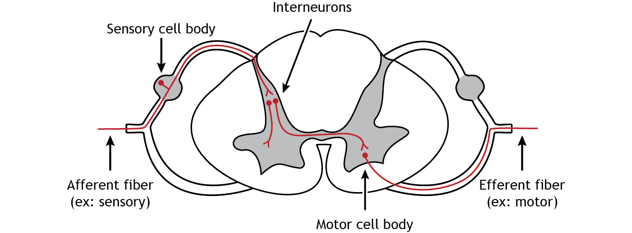
Afferent fibers coming from the periphery through the spinal nerves enter the spinal cord via the dorsal root. The cell bodies of sensory neurons are located in the dorsal root ganglion, and the axons continue into the spinal cord and typically synapse in the dorsal horn. Interneurons are very short neurons that are a communication link between cell types in the spinal cord. They can be either excitatory or inhibitory depending on their role. They can also cross the midline of the spinal cord. The cell bodies of motor neurons that innervate skeletal muscles are located in the ventral horn. The efferent axons of these neurons leave the spinal cord via the ventral root and then enter the spinal nerve on their way to their target tissue.
Key Takeaways
- The brainstem connects the diencephalon to the spinal cord and is responsible for critical body functions
- The cranial nerves enter and exit the central nervous system primarily through the brainstem
- The spinal cord is part of the central nervous system and spinal nerves are part of the peripheral nervous system
- Like the brain, the spinal cord is made up of both white and gray matter
Test Yourself!
