
25 Touch: Central Processing
Receptive Fields and Lateral Inhibition
The receptive fields of the sensory neurons become more complex as information moves up the pathway. We saw in the last lesson that mechanoreceptors have receptive fields that, when touched, activate the neuron. The mechanoreceptors synapse on neurons in the dorsal column, and those neurons have more complex receptive fields. The dorsal column nuclei have receptive fields that are divided into center and surround regions. The center of the receptive field is a result of direct innervation from the mechanoreceptors. If a stimulus touches the skin in the center of a dorsal column neuron’s receptive field, the neuron will increase its firing rate. The center / surround structure is like that of bipolar and ganglion cells in the vision system.
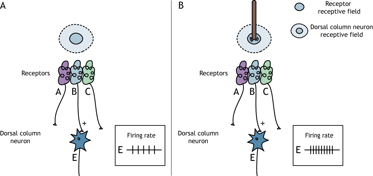
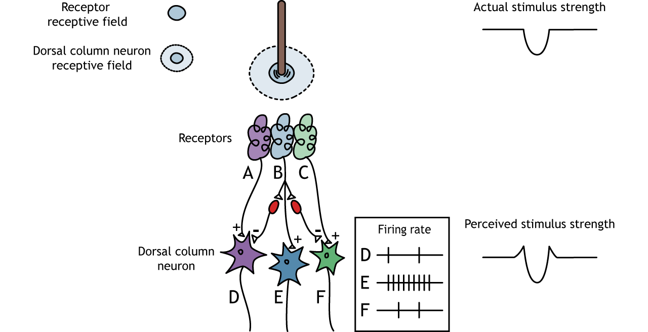
The surround region of the receptive field is a result of indirect communication between the receptor neurons and the dorsal column neurons via inhibitory interneurons. The surround has an inhibitory effect on the dorsal column neuron. If a stimulus touches the skin in the surround of a dorsal column neuron’s receptive field, the neuron will decrease its firing rate.

Lateral inhibition results from overlapping receptive fields and increases perception of stimuli at edges and borders
Lateral Inhibition
The center-surround structure of the receptive field is critical for lateral inhibition to occur. Lateral inhibition is the ability of the sensory systems to enhance the perception of edges of stimuli. At a point or an edge of a stimulus, because of the inhibitory interneurons, the perceived stimulus strength will be enhanced compared to the actual stimulus strength.
Pathway to Brain
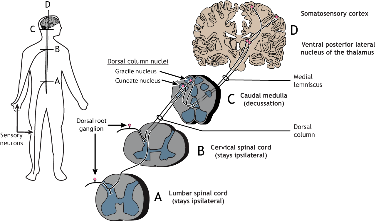
The right side of the brain processes touch on the left side of the body; the left side of the brain processes touch on the right side of the body.
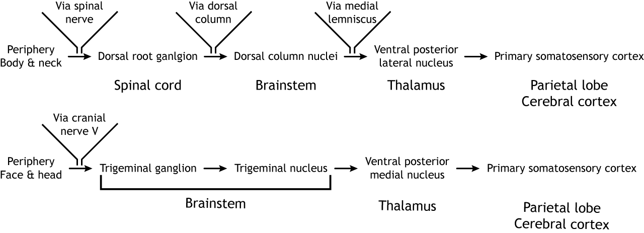
Dorsal Column-Medial Lemniscus Pathway
Primary afferent sensory fibers have their cell bodies located in the dorsal root ganglion, a structure that lies just outside of the spinal cord. The axons of these first-order neurons enter the ipsilateral dorsal side of the spinal cord. Some axon collaterals terminate in the spinal cord and are important for reflexes. The main axon branch ascends the spinal cord toward the brain, via the dorsal column, terminating in the dorsal column nuclei located in the brainstem. The axons of sensory neurons in the lower body remain separate from the axons of sensory neurons in the upper body throughout the pathway. These two populations of neurons synapse in different regions of the brainstem. The lower body axons terminate in the gracile nucleus, whereas the upper body axons terminate in cuneate nucleus. Projections from the second-order neurons in the dorsal column nuclei cross the midline, or decussate, and ascend via a white matter tract called the medial lemniscus. The axons terminate in the ventral posterior lateral nucleus of the thalamus. The thalamic neurons then project to the primary somatosensory cortex located in the postcentral gyrus in the parietal lobe.
View the thalamus using the BrainFacts.org 3D Brain
View the primary somatosensory cortex using the BrainFacts.org 3D Brain
Trigeminal Pathway
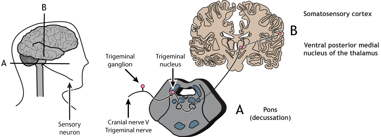
Sensory receptors in the face and head send information to the brain via cranial nerve V, the trigeminal nerve. The first-order neurons have their cell bodies in the trigeminal ganglion, located just outside of the brainstem, and they project to the ipsilateral trigeminal nucleus in the pons. The second-order neurons cross the midline and project up to the ventral posterior medial nucleus of the thalamus. These neurons then send projections to the face region of the somatosensory cortex.
Primary Somatosensory Cortex
Anatomy
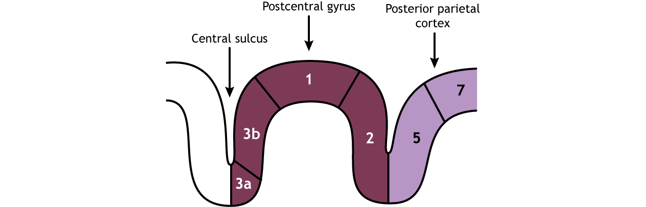
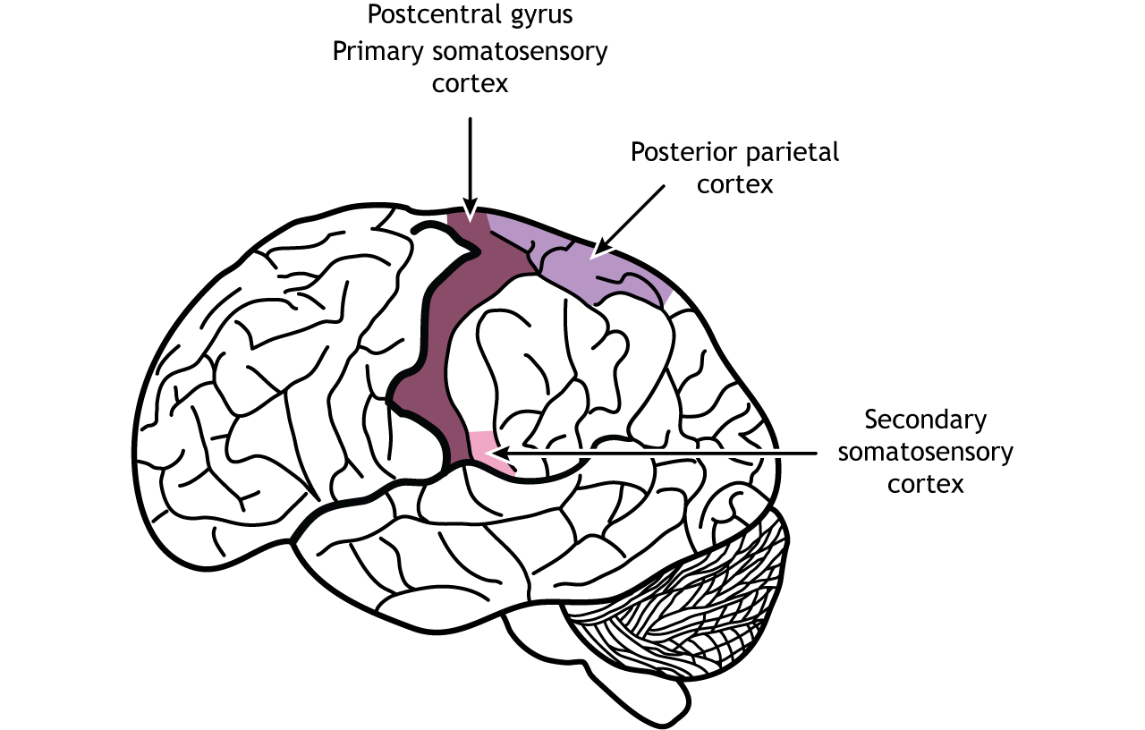
The primary somatosensory cortex is divided into four regions, each with its own input and function: areas 3a, 3b, 1, and 2. Most touch information from mechanoreceptors inputs to region 3b, whereas most proprioceptive information from the muscles inputs to region 3a. These regions then send and receive information from areas 1 and 2. As processing of somatosensory information continues, the stimuli required to activate neurons becomes more complex. For example, area 1 is involved in sensing texture, and area 2 is involved in sensing size and shape of an object. The posterior parietal cortex, an important output region of the somatosensory cortex, lies caudal to the postcentral gyrus; areas 5 and 7 are downstream structures that continue to process touch.
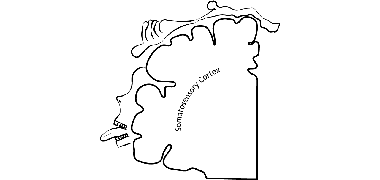
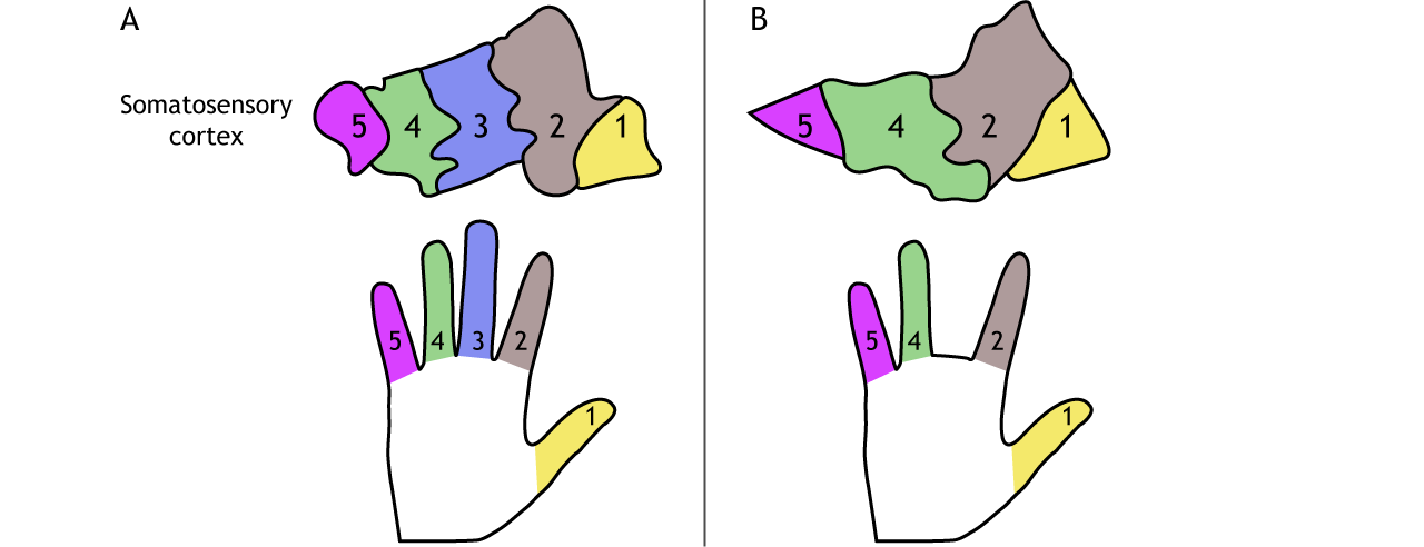
Somatotopic Map
Information from the skin is organized into a map of the body on the primary somatosensory cortex
The receptive fields of each higher order neuron increases in size and complexity, but even cortical neurons are associated with a specific region of the body. Cortical neurons are organized by the region of the body they represent, so neurons that respond to sensation in the fingers are located close to the neurons that respond to sensation in the hand. Remember from above that axons in the dorsal column from the lower body run next to, but remain separate from, the axons from the upper body. This separation, which occurs for all body regions and at all levels of the pathway, creates a somatotopic map of the body in the primary somatosensory cortex. Each area of the somatosensory cortex (Figure 23.7) has its own, but similar, map of the body.
Regions with high receptor density in the skin, and, therefore, fine two-point discrimination, have more cortical space devoted to them. This means that the cortical representation of the body is not true to actual physical proportions. A homunculus is a cartoon representation of what a body would look like if actual body size was proportional to the cortical representation. The hands and lips would be excessively large while the torso, arms, and legs, would be relatively small.
Higher-Level Processing of Touch Information
The primary somatosensory cortex sends projections to other parietal lobe regions for higher-level processing of touch information.
Secondary Somatosensory Cortex
The secondary somatosensory cortex (SII) is located in the inferior parietal lobe, just above the lateral fissure. This region, like the dorsal stream of visual processing, is responsible for object recognition, discerning texture, shape, and size. The SII also has receptive fields that represent bilateral regions of the body, so both hemispheres will be activated by touch on either side of the body. The SII sends projections to the posterior parietal cortex, the premotor cortex, the amygdala, and the hippocampus.
Posterior Parietal Cortex
The posterior parietal cortex recognizes touch characteristics like orientation and movement. It is also important for combining the touch and motor components of actions like grasping. The posterior parietal cortex outputs to the frontal motor cortex.
View the posterior parietal cortex using the BrainFacts.org 3D Brain
Cortical Plasticity
The brain can show plasticity when sensory input changes over time
In adulthood, the brain is plastic, meaning synaptic connections can rearrange under certain conditions. Amputation or loss of a finger, for example, will lead to the associated cortical space to be functionally remapped by input from neighboring regions of the hand. The cortical neurons do not die, they begin to be activated by a different region of the body. Likewise, cortical representation can expand with use or practice. Repeated training of certain fingers can lead to an increase in cortical space mapped to those digits. Cortical plasticity is believed to underly the phenomenon of the perception of phantom limbs after amputation. In these cases, subjects that have lost a region of their body can sometimes still “feel” the missing part.
Key Takeaways
- Receptive fields become more complex as information moves through the touch pathway
- Lateral inhibition enhances edges and borders by affecting the perceived stimulus strength
- Mechanoreceptor afferents synapse in the dorsal column nuclei in the medulla. Information then decussates and synapses in the ventral posterior nucleus of the thalamus before traveling to the primary somatosensory cortex
- Sensory axons from the lower body synapses in the gracile nucleus in the dorsal column
- Sensory axons from the upper body synapses in the cuneate nucleus in the dorsal column
- Information from the neck and body synapse in the ventral posterior lateral nucleus of the thalamus
- Information from the head and face synapse in the ventral posterior medial nucleus of the thalamus
- The primary somatosensory cortex is organized in a somatotopic map
- The cortex is plastic and connections can change with experience
Test Yourself!
- What is the anatomical name of the primary somatosensory cortex?
- After somatosensory information leaves the brainstem, it must relay through which structure before reaching the primary somatosensory cortex?
