
61 HPA Axis
When presented with a stressor, our brain activates the hypothalamic-pituitary-adrenal (HPA) axis, which initiates a hormonal response.
Hypothalamus
The hypothalamus, which sits below the thalamus, integrates information from many regions of the central nervous system and plays a critical role in maintaining homeostasis in the body. The hypothalamus regulates temperature, hunger, thirst, blood volume and pressure, sleep and wakefulness, reproductive functions, and stress and fear responses.
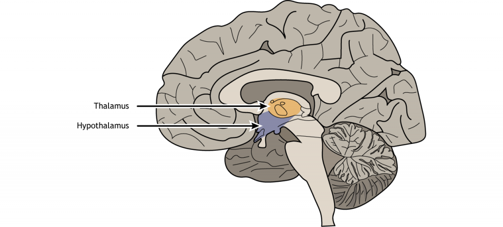
View the hypothalamus using the BrainFacts.org 3D Brain
Pituitary
The hypothalamic regulation of the body’s response to stress is managed via hormone release by the pituitary gland. The pituitary gland is located inferior to the hypothalamus. The pituitary is divided into two lobes, the anterior and the posterior pituitary. These regions are responsible for the release of different hormones and are controlled by the hypothalamus in different ways.
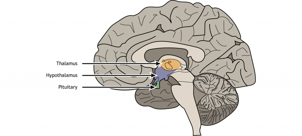
View the pituitary using the BrainFacts.org 3D Brain
Hormone Release
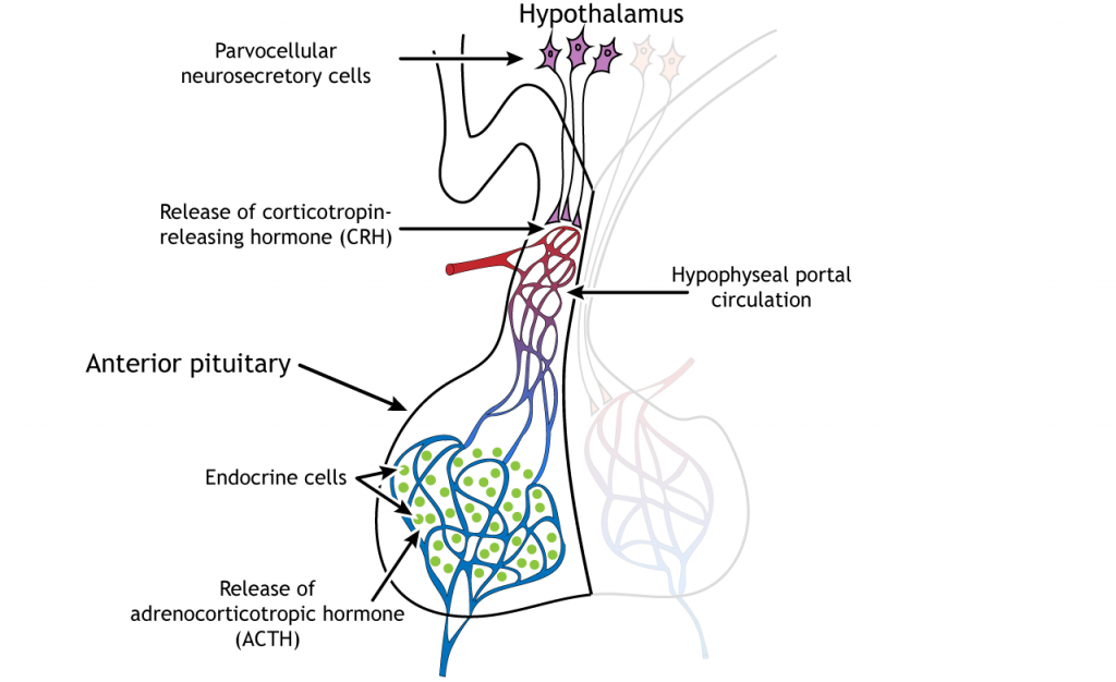
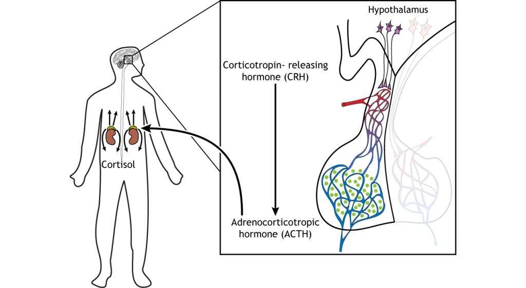
The stress response relies on anterior pituitary function. The hypothalamus contains two types of neurons that secrete hormones into the pituitary: parvocellular neurosecretory cells and magnocellular neurosecretory cells. Parvocellular cells are smaller than the magnocellular neurons (parvus means “small” in Latin). In the HPA axis, the parvocellular neurosecretory cells release a hormone called corticotropin-releasing hormone (CRH) into a specialized capillary system that lies between the hypothalamus and the pituitary called the hypophyseal portal circulation. When CRH reaches the anterior pituitary, it causes the endocrine cells of the pituitary to release adrenocorticotropic hormone (ACTH) into the general circulation.
The ACTH travels through the circulatory system and can act on the adrenal cortex, a gland located on top of the kidney. The adrenal cortex releases cortisol, a glucocorticoid hormone, into the blood stream. Cortisol travels throughout the body and has many effects that prepare the body for either fleeing or fighting the stressor. Promotion of energy use (for a quick escape or for defense) occurs through the release of glucose, the sugar the body uses for energy.
Hormone Action
Cortisol is a steroid hormone; steroid hormones are synthesized from cholesterol and are able to cross the phospholipid bilayer because they are lipid soluble. Glucocorticoid receptors are located in the cytoplasm of many cell types across the body. The receptors dimerize after cortisol binds, and the dimer moves to the nucleus where it can alter DNA transcription.
Animation 61.1. Cortisol can cross the phospholipid bilayer and bind to glucocorticoid receptors. The receptors dimerize, move to the nucleus, and interact with DNA, altering transcription of certain genes. ‘Cortisol Action’ by Casey Henley is licensed under a Creative Commons Attribution Non-Commercial Share-Alike (CC BY-NC-SA) 4.0 International License.
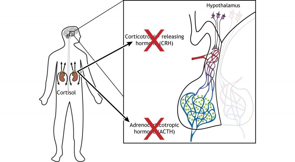
Negative Feedback
Once the stress response has been initiated, and cortisol enters the circulation, cortisol itself is able to act on the hypothalamus and pituitary and inhibit production of CRH and ACTH. This is called a negative feedback loop; the active hormone (cortisol) can shut off its own production. Negative feedback is possible because neurons in the hypothalamus and pituitary express glucocorticoid receptors that are activated by cortisol.
Regulation of the HPA Axis
One area of the brain that regulates the HPA axis is the hippocampus. Activity within the hippocampus normally acts to inhibit activity of the HPA axis. Stressors in the environment inhibit activity of the hippocampus, which in turn disinhibits the HPA axis and allows for cortisol levels to increase. The hippocampus also expresses glucocorticoid receptors that bind to circulating cortisol and act to increase hippocampal activity. This in turn increase inhibition of the HPA axis, another example of negative feedback control within the circuit.

Chronic Stress
While this cortisol response to stress is particularly important in certain situations, like moments of danger, chronic stress is an unhealthy scenario which can put people at risk for heart disease and other illnesses. Chronic stress can cause structural and functional changes, like cell death or alterations in the dendritic arbor, within the cortical regions that play a role in control of the HPA axis and the hippocampus due to long-lasting exposure to cortisol.
Key Takeaways
- The hypothalamus directly controls the stress response by controlling hormone release from the anterior pituitary.
- The hypothalamus releases corticotropin-releasing hormone (CRH).
- The anterior pituitary releases adrenocorticotropic hormone (ACTH).
- The adrenal cortex releases cortisol.
- Cortisol binds to receptors and alters DNA transcription.
- Cortisol can shut off its own production via a negative feedback loop.
Attributions
Portions of this chapter were remixed and revised from the following sources:
- Foundations of Neuroscience by Casey Henley. The original work is licensed under a Creative Commons Attribution-NonCommercial-ShareAlike 4.0 International License
Media Attributions
- Hypothalamus Sagittal © Casey Henley is licensed under a CC BY-NC-SA (Attribution NonCommercial ShareAlike) license
- Hypothalamus Pituitary © Casey Henley is licensed under a CC BY-NC-SA (Attribution NonCommercial ShareAlike) license
- CRH and ACTH Release © Casey Henley is licensed under a CC BY-NC-SA (Attribution NonCommercial ShareAlike) license
- HPA Axis © Casey Henley is licensed under a CC BY-NC-SA (Attribution NonCommercial ShareAlike) license
- Cortisol Feedback © Casey Henley is licensed under a CC BY-NC-SA (Attribution NonCommercial ShareAlike) license
- HPA axis with hippocampus © Valerie Hedges is licensed under a CC BY-NC-SA (Attribution NonCommercial ShareAlike) license
Below; toward the feet
