
21 Anatomical Terminology
The nervous system is one of the most complex systems that we know of. Parts of this system malfunction frequently, and the results are a wide range of neurological disorders that affect humans, from injury to genetic disorders. The gross anatomy of the nervous system is an important foundation to the studies of other aspects of neuroscience. This chapter covers some of the major anatomical structures in the nervous system.
Divisions of the Nervous System
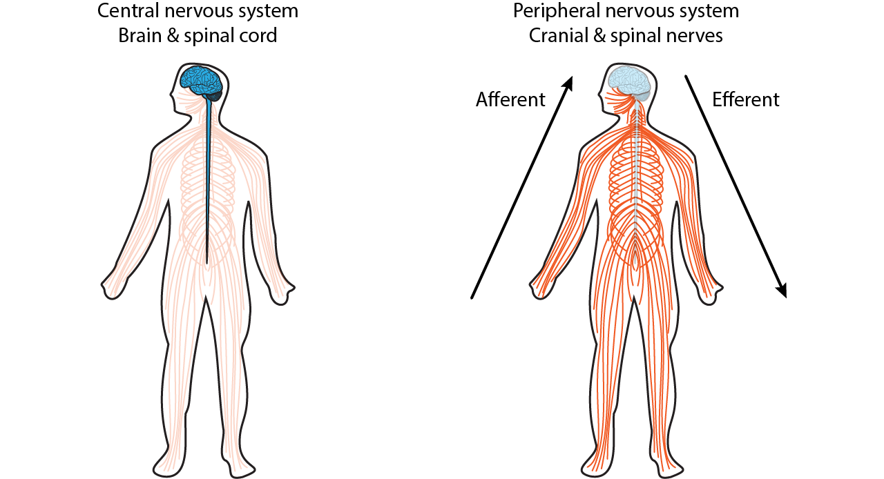
Broadly speaking, the nervous system can be divided into two main categories: The central nervous system (CNS) and the peripheral nervous system (PNS). Simply put, the CNS is the brain and spinal cord, while the PNS is all the other nerve cells in the body in the periphery. The two systems are not isolated from each other; information passes rapidly between the PNS and the CNS, and vice versa. When a signal that originates in the PNS moves to the CNS, we sometimes say that the signal is incoming or ascending, while the CNS to PNS direction is outgoing or descending. Information that arrives into the CNS is also called an afferent signal, while information leaving the CNS is an efferent signal. These two terms are frequently confused, but you can use the knowledge of other words that start with “e” to remember that an “exit” or an “escape” is something that moves away for ‘efferent’, and “arrive” for ‘afferent’. Alternatively, an efferent signal is something that has an effect on the outside world, while an afferent signal affects the person.
Anatomically, the CNS consists of two organs, the brain and the spinal cord. The brain is the main organ where movement originates, where thoughts and plans develop, and where consciousness is housed. The brain is what pushes us to act on our drives and desires, where language begins, and where memories are stored. The intact adult brain weighs about 1.5 kg (3 pounds), which is barely 2% of total body weight. Despite this relatively small size, it is extremely power hungry, and uses up about one-fifth of the body’s total energy expenditure.
Anatomical Terms
When talking about parts of the brain, it is helpful to have a set of directional terms that can describe the location of various anatomical structures unambiguously.
Familiarity with the terminology used to describe location and relationships within the nervous system is critical as we move forward into examining brain systems.
Directional Terms
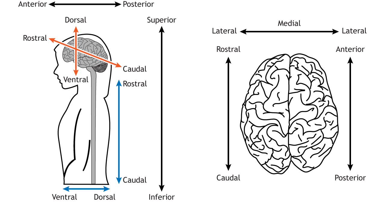
Directional terms are used to locate one structure, usually in relation to another structure. Some terms, like dorsal or ventral, are relative to the axis of the central nervous system, so the direction these terms define changes if used for brain regions versus other body regions. Other terms, like superior or inferior, keep their meaning across the entire body.
- Anterior: In front of; toward the face
- Posterior: Behind; toward the back
- Superior: Above; toward the head
- Inferior: Below; toward the feet
- Medial: Toward the middle
- Lateral: Toward the edge
- Dorsal: Toward the top of the brain or the back of the spinal cord
- Ventral: Toward the bottom of the brain or the front of the spinal cord
- Rostral: Toward the front of the brain or the top of the spinal cord
- Caudal: Toward the back of the brain or the bottom of the spinal cord
Often anterior and rostral are used interchangeably and posterior and caudal are used interchangeably.
Neuroanatomists use all of these terms to describe the relationship of one structure to another. For example, we’ll see in Chapter 22 that introduces the four lobes of the brain, the frontal lobe is anterior or rostral to the parietal lobe, and the parietal lobe is dorsal to the temporal lobe. These anatomical words can also be combined to subdivide complex brain regions. For example, a structure called the thalamus has many small subsections, such as the dorsomedial nucleus or the ventropostero-lateral nucleus. Naming structures with this anatomical language is useful in identifying where they are located in a brain scan or autopsy, but these words tell us nothing about function.
Visualizing the Brain: Anatomical Planes
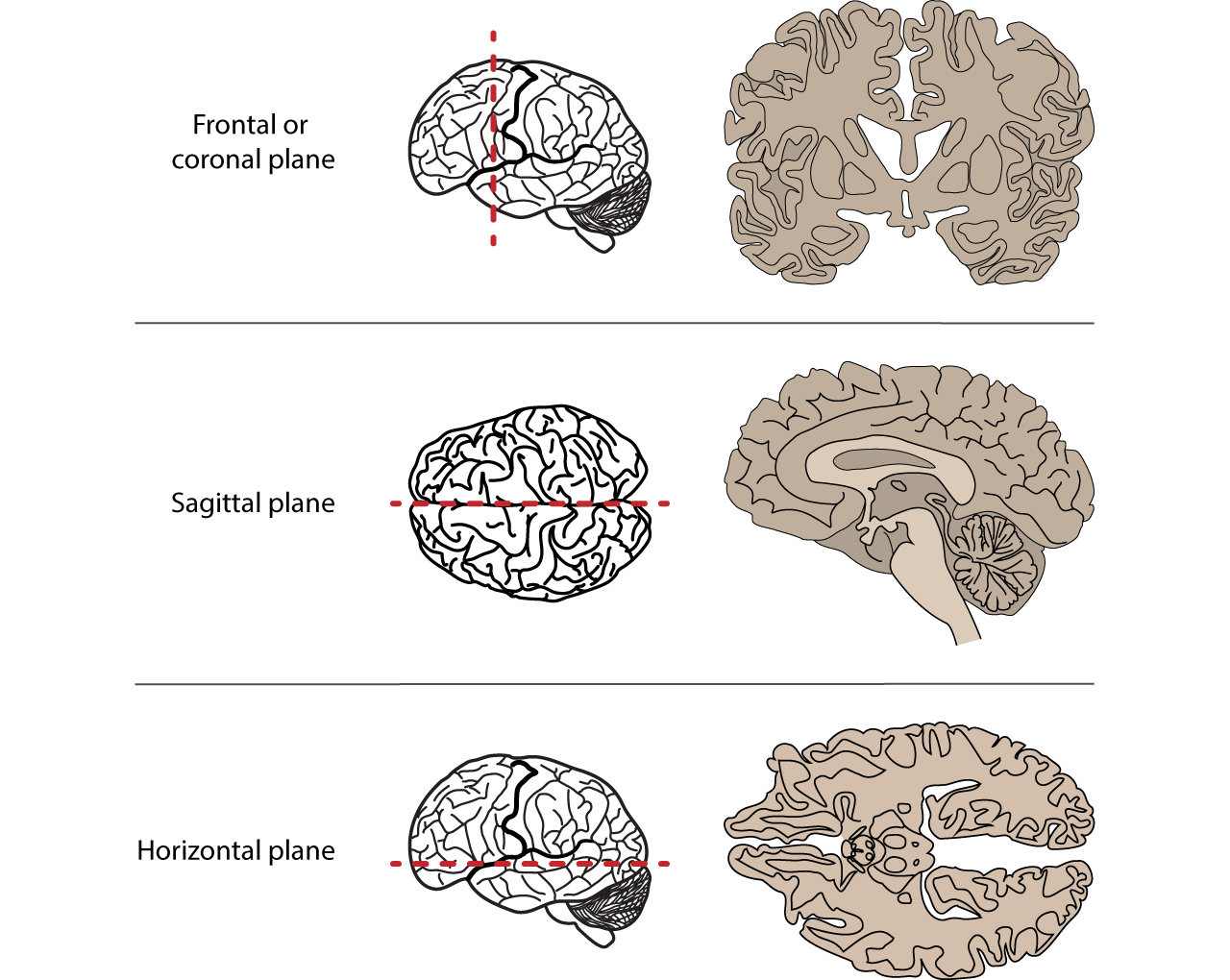
As a three-dimensional structure, the brain can be sectioned, or cut, for visualization or analysis in several ways. Here, we will describe the three main orientations, each at right angles to the others.
- The frontal or coronal plane is a vertical plane in a medial to lateral direction, dividing objects into front and back pieces.
- The sagittal plane is also a vertical plane but in a rostral-caudal direction, meaning it divides objects into right and left regions.
- Finally, the horizontal plane divides objects into top and bottom regions.
Gray Matter and White Matter
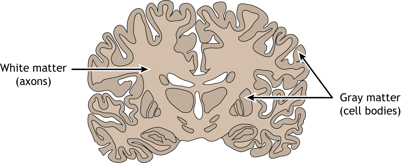
In a sliced section of the brain, you might notice that brain tissue has different colored areas. Some brain tissue is pale and almost white, and these areas are described as white matter. Generally, white matter represents pathways of communication. White matter regions are comprised of axons. It appears white due to the myelin sheath on the axons. Other sections of brain tissue have a darker pink/gray color, appropriately called gray matter. These areas are usually dense with cell bodies and dendrites. Gray matter is the location of most synapses.
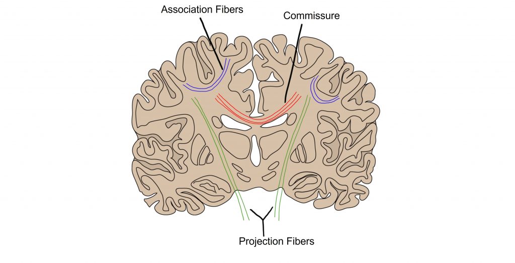
White Matter Tracts
The brain has two very similar halves, the left and right hemisphere. Oftentimes, in the neurotypical individual, information passes between both hemispheres rapidly: what one hemisphere senses or learns, so does the other hemisphere. It is the white matter tracts that allow for this transfer of information. When a white matter pathway crosses from one side of the body to another, we call it a decussation.
Commissures are white matter tracts that connect gray matter between the left and right hemispheres of the brain. The main commissure that allows for the passage of information between the two hemispheres is called the corpus callosum.
Association fibers are white matter tracts that connect different bits of gray matter within the same hemisphere of the brain. These connections do not cross the midline.
Projection fibers are white matter tracts that connect the hemispheres of the brain with lower brain structures or with the spinal cord. They “project” from the cortex to the lower brain structures or spinal cord.
Key Takeaways
- Anatomical terminology is critical for determining neurological landmarks and discussing the anatomy of the nervous system
Test Yourself!
Attributions
Portions of this chapter were remixed and revised from the following sources:
- Foundations of Neuroscience by Casey Henley. The original work is licensed under a Creative Commons Attribution-NonCommercial-ShareAlike 4.0 International License
- Open Neuroscience Initiative by Austin Lim. The original work is licensed under a Creative Commons Attribution-NonCommercial 4.0 International License.
Media Attributions
- White Matter Connections © Valerie Hedges is licensed under a CC BY-NC-SA (Attribution NonCommercial ShareAlike) license
The brain and spinal cord
All nervous system components that are NOT the brain and spinal cord
traveling towards the CNS
traveling from the CNS to the body
in front of; toward the face
Behind; toward the back
Above; toward the head
Below; toward the feet
Toward the middle
Toward the edge
Toward the top of the brain or the back of the spinal cord
Toward the bottom of the brain or the front of the spinal cord
Toward the front of the brain or the top of the spinal cord
Toward the back of the brain or the bottom of the spinal cord
