
41 Spinal Reflexes
Reflexes
Reflexes are involuntary motor responses that are performed automatically and independent of brain signals (although some can be suppressed voluntarily, with extra effort). Reflexes involve very simple circuits, sometimes consisting of only two populations of neurons: Sensory information comes in from the periphery, synapsing onto motor neurons in the brainstem or spinal cord. Reflexes can take place as quickly as 1/100th of a second!
Stretch Reflex
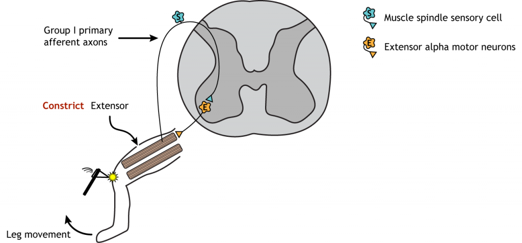
When a muscle is stretched, this stretches the associated muscle spindle, which activates a 1a sensory afferent neuron that has its cell body located in the dorsal root ganglion. The sensory axon enters the dorsal side of the spinal cord where it makes two synapses:
- A direct synapse onto a motor neuron in the spinal cord gray matter. The axon of the motor neuron exits the spinal cord and innervates the stretched muscle, causing it to contract.
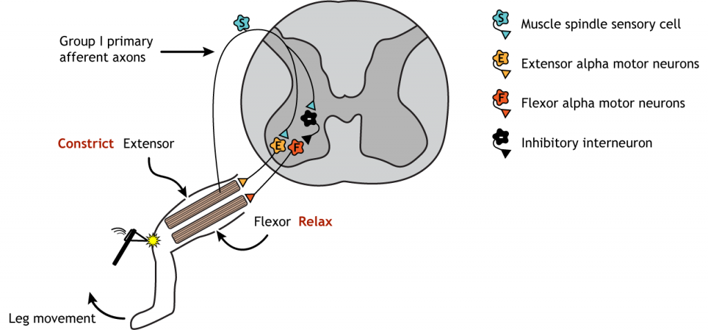
- A synapse onto an inhibitory interneuron that then synapses onto a separate motor neuron that innervates the antagonistic muscle.
The myotatic reflex, also called the patellar, or knee-jerk reflex, is an example of a stretch reflex and occurs in response to activation of the muscle spindle stretch receptors. This reflex is commonly tested at a doctor’s visit when the doctor taps your knee with a little hammer. This usually results in the lower leg kicking up slightly. The synaptic communication for this reflex takes place completely within the spinal cord and requires no input from the brain.
The knee is tapped on the tendon that connects to the quadriceps muscle. The tapping of the tendon stretches the tendon and as a result causes the quadriceps muscle to stretch, activating the stretch receptors. Sensory information travels to the dorsal horn of the spinal cord where it synapses on alpha motor neurons that innervate the quadriceps muscle. Activation of the motor neurons contracts the quadriceps, extending the lower leg. This is called a monosynaptic communication because there is only one synapse between the sensory input and the motor output. The reflexive kick is controlled at the level of the spinal cord and cannot be intentionally suppressed by descending motor pathways no matter how hard you concentrate.
The sensory neurons also synapse on interneurons within the spinal cord that are inhibitory (this means that these neurons make and release GABA). These inhibitory interneurons then synapse on alpha motor neurons that innervate the hamstring, the antagonistic flexor muscle to the quadriceps. When these motor neurons are inhibited, the hamstring muscle relaxes, allowing the contraction of the quadriceps to occur with more ease. This is called reciprocal inhibition.
Withdrawal (Flexor) Reflex
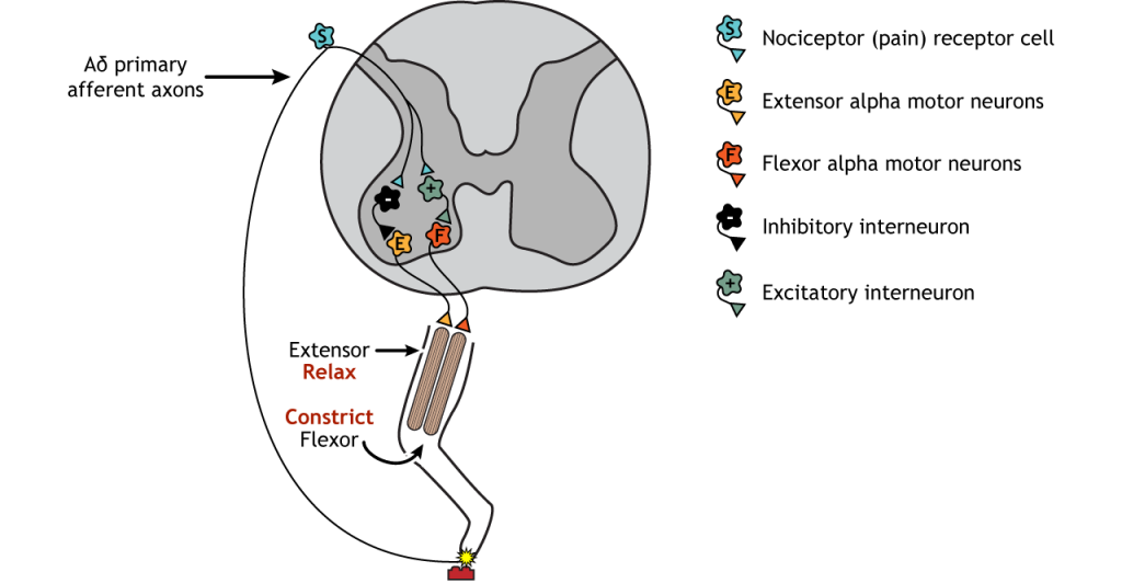
A similar process can be seen in the withdrawal reflex. In this case, instead of an extension, the muscles lead to muscle flexion in response to a stimulus. If, for example, you step on something painful, the reflex will be to lift the injured foot. The sensory information that initiates this reflex is activation of pain receptors, or nociceptors. Like with the stretch reflex, the sensory information enters the spinal cord at the dorsal horn. Unlike the stretch reflex, the withdrawal reflex is a polysynaptic reflex, meaning interneurons are present between the sensory neurons and the motor neurons. Excitatory interneurons communicate with the alpha motor neurons of the flexor muscle, whereas inhibitory interneurons communicate with the alpha motor neurons of the extensor muscle. The behavioral response is flexing of the leg upward (the opposite action of the stretch reflex).
Crossed-Extensor Reflex
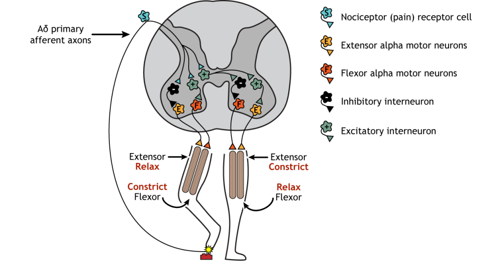
Running in parallel to the withdrawal reflex is the crossed-extensor reflex. If you step on something sharp and lift that leg, your other leg needs to be able to support your weight shift, or you would fall. This is accomplished by interneurons that cross the midline of the spinal cord and communicate with motor neurons on the contralateral side of the body. The painful sensory information that initiated the withdrawal reflex also initiates the crossed-extensor reflex. In addition to the ipsilateral interneurons active in the withdrawal reflex, the sensory axons also synapse on excitatory interneurons that cross the midline. These interneurons then synapse on excitatory interneurons that activate the alpha motor neurons of the extensor muscle and inhibitory interneurons that inhibit the alpha motor neurons of the flexor muscle (the opposite configuration to the withdrawal reflex). This leads to the leg extending, providing a stable base for the weight shift.
Key Takeaways
- Control of reflexes occurs within the spinal cord and input from the brain is not needed.
Test Yourself!
Attributions
Portions of this chapter were remixed and revised from the following sources:
- Foundations of Neuroscience by Casey Henley. The original work is licensed under a Creative Commons Attribution-NonCommercial-ShareAlike 4.0 International License
- Open Neuroscience Initiative by Austin Lim. The original work is licensed under a Creative Commons Attribution-NonCommercial 4.0 International License.
Media Attributions
- Stretch Reflex Extensor © Casey Henley adapted by Valerie Hedges is licensed under a CC BY-NC-SA (Attribution NonCommercial ShareAlike) license
- Stretch Reflex © Casey Henley adapted by Valerie Hedges is licensed under a CC BY-NC-SA (Attribution NonCommercial ShareAlike) license
- Withdrawal Reflex © Casey Henley adapted by Valerie Hedges is licensed under a CC BY-NC-SA (Attribution NonCommercial ShareAlike) license
- Crossed Extensor Reflex © Casey Henley adapted by Valerie Hedges is licensed under a CC BY-NC-SA (Attribution NonCommercial ShareAlike) license
Involuntary motor responses that are performed automatically and independent of brain signals
Proprioceptor that communicates information about muscle length
occurs on the same side of the body
