
26 Spinal Cord Structure
It is sometimes easy to think of neuroscience as a focused study of the brain: How does activity of the brain contribute to behavior? In what ways does the brain change in disease? Why do the cells of the brain behave the way they do?
The truth is there are many parts of the body that also fall under the broad study of neuroscience. For example, the automatic kneejerk reflex that a clinician examines when they tap on your patellar tendon is a test of the nervous system. The reflex is driven by sensory neurons that detect muscle stretch, motor neurons that cause the kicking response, and interneurons that prevent the opposing muscle from acting. We have neural circuits that provoke changes in the activity of our internal organs, from the beating of our heart to the digestion of food, and the study of these systems is certainly part of neuroscience as well.
Moving posterior from the brainstem is the other organ of the central nervous system: a long, thin structure of nervous tissue called the spinal cord. It functions to carry information both upwards towards the brain and downwards towards the body’s other organs and muscles. It can also process sensations and form an appropriate motor response in the absence of brain input.
Spinal Column Anatomy
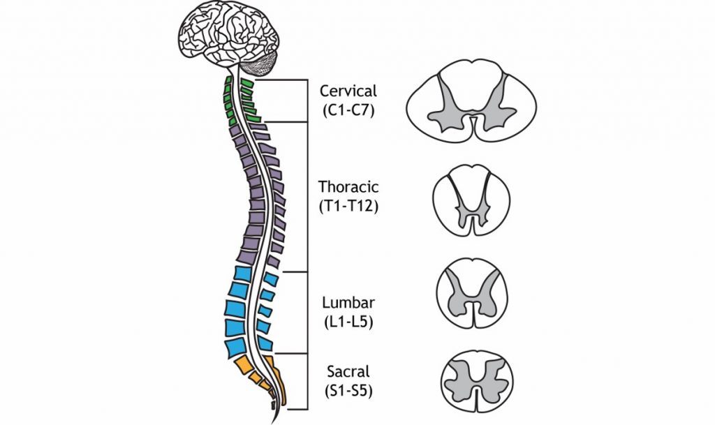
The spinal cord begins at the base of the brainstem and runs down to the small of your back, giving it a length around 44 cm (17.5 inches). The spinal cord is housed within a series of bones, called the vertebral column. Although the spinal cord itself is continuous, it can be divided based on the overlying vertebrae. A combination of a letter and a number is used to identify each section of the spinal cord; the letter corresponds to the vertebral section and the number refers to the number of bones down from the previous section (the smaller numbers are more anterior, larger numbers more posterior). The diameter and shape of the spinal cord changes over the length of vertebral column, a result of the function of the spinal nerves. For example, the region where motor neurons are located (ventral horn) is larger in the cervical region of the spinal cord compared to other regions of the spinal cord with minimal motor output.
Branching off from each section of the spinal cord are two pairs of nerves, the afferent (incoming to the CNS) sensory nerve roots, which branch from the dorsal side of the spinal cord, and the efferent (outgoing from the CNS) motor nerve roots, which branch from the ventral side of the spinal cord. These two branches meet and extend away from the spinal cord. After merging, they are called the spinal nerves.
The vertebral column is divided into four main regions: cervical, thoracic, lumbar, and sacral. The spinal cord and spinal nerves that enter and exit the vertebral column are divided into these regions as well. Moving from anterior (top) to posterior (bottom), the four regions of the spinal cord are:
- Cervical. The cervical region corresponds to C1 through C7. Nerves that exit through the cervical region innervate the muscles in the neck, shoulders, arms, and hands. Afferent nerves detect somatosensory inputs from these same areas. Sections C3 through C5 innervate the diaphragm, so an injury at this level or higher can quickly lead to death since the person may stop breathing. The spinal cord is at the widest diameter at the cervical area, as it has a swelling that corresponds to the many inputs and outputs to the arms.
- Thoracic. The thoracic region corresponds to T1 through T12. These regions innervate the middle trunk area, the intercostal muscles between the ribs, and abdominal muscles. Branches of the spinal nerves in the thoracic areas are responsible for changing the activity of the various internal organs during a fight-or-flight response (more on the autonomic nervous system in Chapter 27).
- Lumbar. The lumbar region corresponds to L1 through L5. These pathways carry motor command information to the hips, thighs, and knees. Afferent lumbar inputs detect sensory information from the ventral side of the legs, such as the top of the thigh or the shin bone. As in the cervical region, the lumbar region has a swelling that increases the diameter of this section of spinal cord compared to the thoracic or sacral areas.
- Sacral. At the posterior-most end of the spinal cord is the sacral region, which corresponds to S1 through S5. Sacral spinal nerves control flexing of the toes. These nerves detect sensory information around the genital organs and the dorsal aspects of the legs, like the buttocks and the back of the thighs. There are also parasympathetic nerves that come from the sacral region and these innervate the colon, bladder, and genital organs (more on the autonomic nervous system in Chapter 27). Since information must pass through the anterior regions of the spinal cord to reach the posterior parts of the body, the more anterior an injury, the more parts of the body that are affected.
From L2 through S5, the spinal cord exists as the cauda equina, meaning “horse’s tail” due to the appearance of the cord as individual nerves that branch from the main spinal cord. These nerves then innervate the pelvis area and lower limbs. This area of the spinal cord is where spinal tap and epidural procedures take place due to the decreased risk of spinal injury.

The spinal cord is part of the central nervous system, but the fibers that leave and enter the spinal cord are located in the peripheral nervous system. These spinal nerves can then extend to or from target tissues throughout the body.
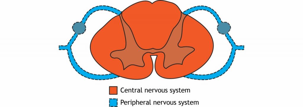
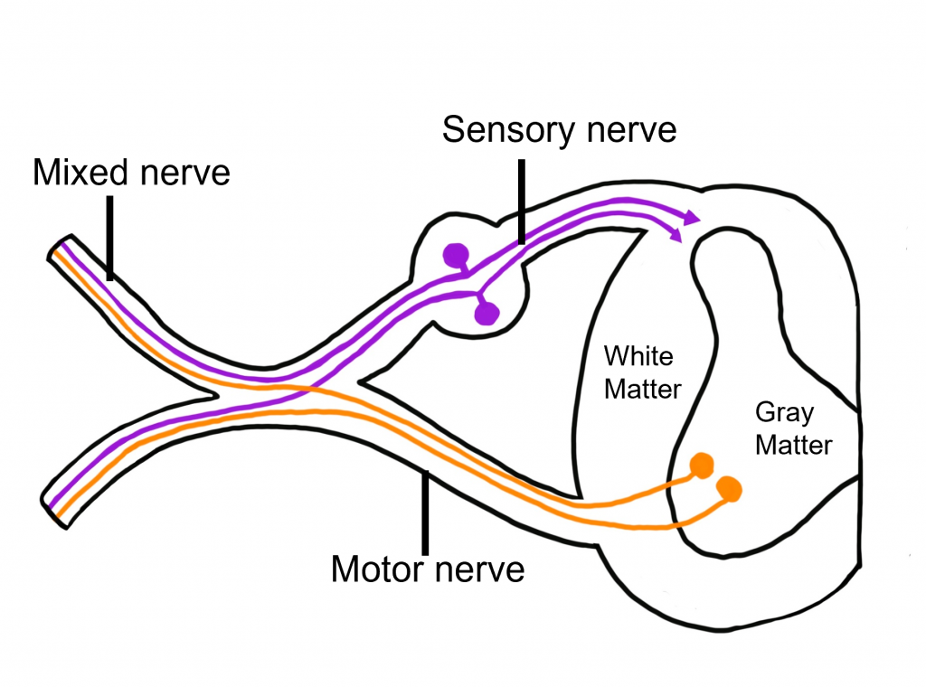
Nerves are collections of neuron axons found within the peripheral nervous system and can be classified as sensory, motor, or mixed nerves. The dorsal root is an example of a sensory (afferent) nerve that is responsible for carrying information toward the central nervous system. The ventral root is an example of a motor (efferent) nerve that is responsible for carrying information away from the central nervous system. Most nerves in the body are classified as mixed nerves that contain both sensory and motor fibers.
Cross Section Anatomy of the Spinal Cord
Like the brain, the spinal cord is also made up of regions of white matter and gray matter. White matter regions are comprised of axons. It appears white due to the myelin sheath on the axons. Gray matter regions are comprised of cell bodies and dendrites. Gray matter is the location of most synapses.
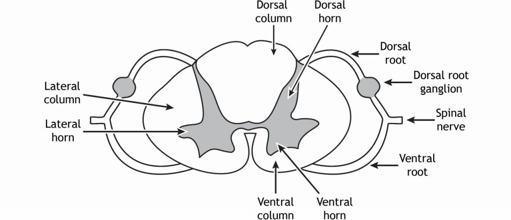
In cross section, the gray matter of the spinal cord is found medially, and the white matter is found laterally. When referring to the spinal cord, we will typically use the directional terms “dorsal” and “ventral”. By convention, when looking a cross section of the spinal cord (a horizontal cut through the cord), the dorsal portion of the spinal cord will be located at the top of the image and the ventral portion of the spinal cord will be located at the bottom of the image. There are a few structures to be aware of when examining the spinal cord in cross section.
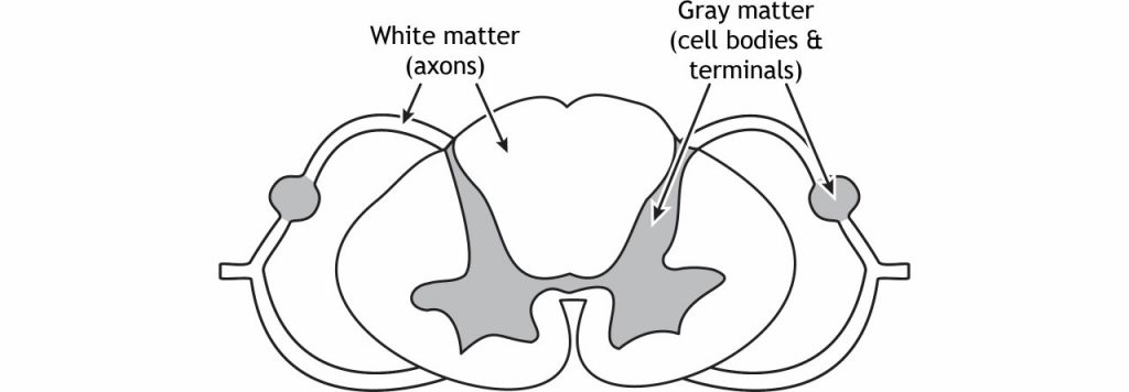
The white matter in the spinal cord is divided into structures called columns because the axons in these regions are either ascending toward the brain or descending toward the appropriate spinal nerve. The dorsal column is on the dorsal or posterior side of the spinal cord, the ventral horn is on the ventral or anterior side of the spinal cord, and the lateral column lies between them. The gray matter is likewise divided into regions called horns. The dorsal horn is the location of sensory synapses, the ventral horn is the location of motor neuron cell bodies, and the lateral horn is the location of cell bodies of the autonomic nervous system. The dorsal root and ventral root consist of the axons of afferent (dorsal) and efferent (ventral) fibers. They combine to form the spinal nerves. Sensory neuron cell bodies are located in the dorsal root ganglion, a gray matter region of the dorsal root.
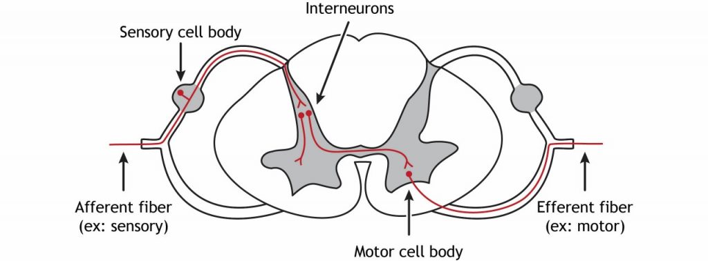
All somatosensory receptor neurons have their cell bodies located in the dorsal root ganglion; a structure found just outside the dorsal aspect of the spinal cord. The receptor neurons (also called primary afferent fibers) of the somatosensory system are bipolar neurons, meaning they have one process from the cell body that splits into two branches. Afferent fibers coming from the periphery through the spinal nerves enter the spinal cord via the dorsal root. The cell bodies of sensory neurons are located in the dorsal root ganglion. The axons continue into the spinal cord and typically synapse in the dorsal horn. Interneurons are very short neurons that are a communication link between cell types in the spinal cord. They can be either excitatory or inhibitory depending on their role. They can also cross the midline of the spinal cord. The cell bodies of motor neurons that innervate skeletal muscles are located in the ventral horn. The efferent axons of these neurons leave the spinal cord via the ventral root and then enter the spinal nerve on their way to their target tissue.
The ventral portion of the spinal cord is concerned with motor output, or efferent signals. Muscle fibers are innervated by alpha motor neurons that have their cell bodies in the ventral horn of the spinal cord. Their axons leave the spinal cord via the ventral roots and travel to the muscle via efferent peripheral spinal nerves.
In summary, sensory information is concerned with the dorsal portion of the spinal cord whereas motor information is concerned with the ventral portion of the spinal cord.
Key Takeaways
Type your key takeaways here.
- There are four regions of the spinal cord
- There are gray matter and white matter areas of the spinal cord
- Sensory information enters into the spinal cord via the dorsal root ganglion to the dorsal horn
- Sensory cell bodies are located within the dorsal root ganglion
- Motor information exits the spinal cord from the ventral horn then through the ventral root out to the body
Test Yourself!
Attributions
Portions of this chapter were remixed and revised from the following sources:
- Foundations of Neuroscience by Casey Henley. The original work is licensed under a Creative Commons Attribution-NonCommercial-ShareAlike 4.0 International License
- Open Neuroscience Initiative by Austin Lim. The original work is licensed under a Creative Commons Attribution-NonCommercial 4.0 International License.
Media Attributions
- Veretbral Column
- Cauda equina © John A Beal adapted by Valerie Hedges is licensed under a CC BY (Attribution) license
- Spinal Cord CNS and PNS
- Nerve Classifications
- Spinal Cord White and Gray Matter
- Spinal Cord Anatomy
- Spinal Cord Fibers
traveling towards the CNS
traveling from the CNS to the body
in front of; toward the face
Behind; toward the back
A bundle of axons in the peripheral nervous system
Toward the top of the brain or the back of the spinal cord
Toward the bottom of the brain or the front of the spinal cord
