
43 Planning of Movement
All voluntary (or non-reflexive) movements begin as signals in the brain. Specifically, the neurons involved in motor control are primarily found in the frontal lobe of the telencephalon, which includes areas such as primary motor cortex (or M1), premotor cortex, and supplemental motor areas. The posterior parietal cortex also contributes to movement. Through this section, we will walk through the brain processes leading to voluntary motor action.
There are a number of steps that must take place for voluntary movement to occur. Assessment of the surrounding environment and the body’s location in space, followed by determining what action is appropriate, and then initiating that action.
For example, after work, you sit down on the couch to watch one episode of your favorite show. As the end credits appear, you realize it is now time to head to your study space and start working on class. To do this, you need to leave the couch, grab your computer from the table, get your coffee from the kitchen and head to a different room. All of these voluntary movements take a great deal of processing by the brain. You must assess your surrounding environment and your body’s location in it, determine which actions need to be completed, and then actually initiate those actions. In this chapter we will focus on how the planning of voluntary movement occurs.
Cortical Anatomy
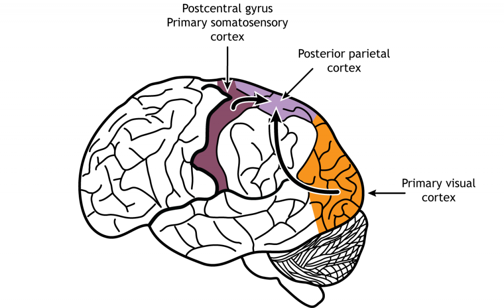
Much of the cortex is actually involved in the planning of voluntary movement. Sensory information, particularly the dorsal stream of the visual and somatosensory pathways, are processed in the posterior parietal lobe where visual, tactile, and proprioceptive information are integrated. The posterior parietal cortex is largely concerned with integrating somatosensory with visual information and determining an appropriate motor action. For example, if you were planning to get up from your seat to walk across the room, the posterior parietal cortex would take in the somatosensory proprioceptive information about how your body is positioned, and the visual information from the objects in the room to avoid running into them (recall the dorsal stream pathway).
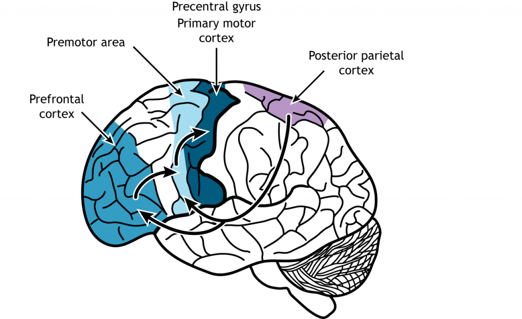
With respect to motor control, the prefrontal cortex (PFC), located in the front of the brain in the frontal lobe initiates the long-term planning or cognitive aspects of movements. For example, consider the motor actions related to brushing your teeth. PFC signals are more akin to “brushing is good for my hygiene and health”, rather than “move my arm and open my mouth.” PFC also helps determine if some motor action is appropriate for the specific situation. Think of a behavioral test where you are given two clickers, one to hold in each hand. You are told that when the experimenter shows you a green item, you should click with the right button. After repeating this behavior multiple times, you are told to switch – now, when you see a green item, you need to click the left button. In this experiment, PFC is responsible for deciding which motor pattern (left button or right button press) is appropriate in response to the stimulus. PFC also works to weigh the consequences of motor actions and makes updates about future motor actions in similar or different circumstances. The exact same motor action produces different results depending on the specific situation, and PFC contributes to evaluating and predicting outcomes.
The premotor area lies just anterior to the primary motor cortex (M1) (M1 is just anterior to the central sulcus). The premotor area helps plan and organize movement and makes decisions about which actions should be used for a situation. The premotor area modulates motor output, and generally activates prior to motor activity.
View the primary motor cortex using the BrainFacts.org 3D Brain
View the premotor cortex using the BrainFacts.org 3D Brain
Role of Premotor Area
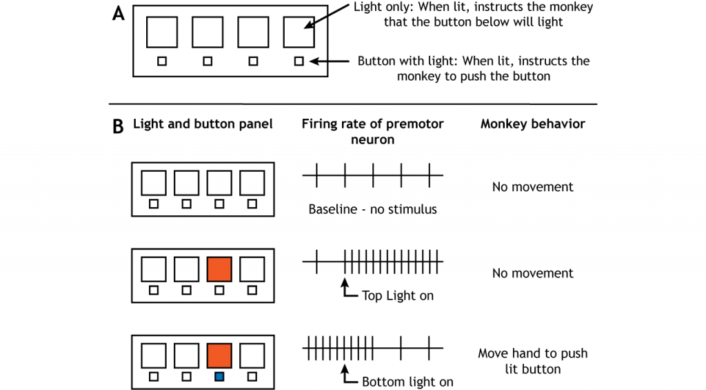
The premotor regions do send some axons directly to lower motor neurons in the spinal cord using the same pathways as the motor cortex (see Execution of Movement chapter). However, the premotor cortex also plays an important role in the planning of movement. Two experimental designs have demonstrated this role. Monkeys were trained on a panel that had one set of lights in a row on top and one set of buttons that could also light up in a row on the bottom. The monkeys would watch for a top row light to turn on. This would indicate that within a few seconds, the button directly below would light up. When the button turned on, the monkeys were supposed to push the button.
Therefore, there were two light triggers in the experiment. The first required no motor movement from the monkey but did give the monkey information about where a motor movement would be needed in the near future. The second required the monkey to move to push the button. When brain activity was measured during this study, neurons in the premotor cortex became active when the first light trigger turned on, well before any movement actually took place (Weinrich and Wise, 1928).
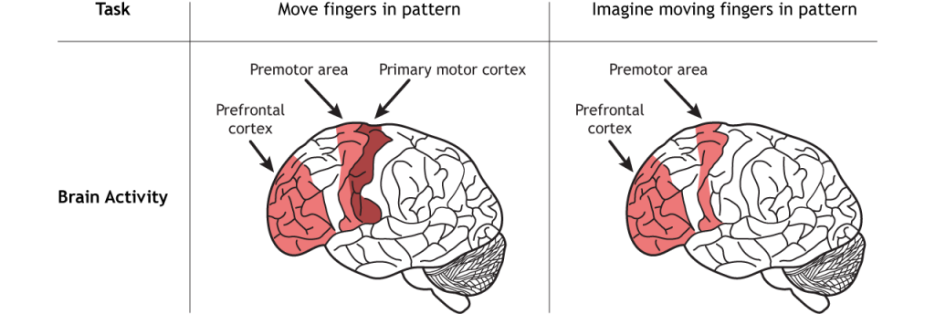
In another experiment, people were trained to move their fingers in a specific pattern. Cerebral blood flow was then measured when they repeated the finger pattern and when they only imagined repeating the finger pattern. When the movement was only imagined and not actually executed, the premotor regions along with parts of the prefrontal cortex were activated (Roland, et al, 1980).
These studies show that the premotor cortex is active prior to the execution of movement, indicating that it plays an important role in the planning of movement. The posterior parietal, prefrontal, and premotor regions, though, also communicate with a subcortical region called the basal ganglia to fully construct the movement plan. The basal ganglia are covered in a future chapter.
Mirror Neurons
There is debate about the existence of a special population of cells in the premotor area called mirror neurons. These are cells that are active during a movement, but also when that same movement is observed in another animal or person. Proponents argue that these neurons are involved in learning behaviors and for understanding the behaviors of others.
Key Takeaways
- Sensory information is processed in the posterior parietal before being sent to motor regions of the brain.
- The prefrontal cortex and premotor cortex are critical for creating a movement plan.
- Mirror neurons
Test Yourself!
References
Roland PE, Larsen B, Lassen NA, Skinhøj E. Supplementary motor area and other cortical areas in organization of voluntary movements in man. J Neurophysiol. 1980 Jan;43(1):118-36. doi: 10.1152/jn.1980.43.1.118. PMID: 7351547.
Weinrich M, Wise SP. The premotor cortex of the monkey. J Neurosci. 1982 Sep;2(9):1329-45. doi: 10.1523/JNEUROSCI.02-09-01329.1982. PMID: 7119878; PMCID: PMC6564318.
Attributions
Portions of this chapter were remixed and revised from the following sources:
- Foundations of Neuroscience by Casey Henley. The original work is licensed under a Creative Commons Attribution-NonCommercial-ShareAlike 4.0 International License
- Open Neuroscience Initiative by Austin Lim. The original work is licensed under a Creative Commons Attribution-NonCommercial 4.0 International License.
Media Attributions
- Posterior Parietal © Casey Henley adapted by Valerie Hedges is licensed under a CC BY-NC-SA (Attribution NonCommercial ShareAlike) license
- Motor Regions © Casey Henley adapted by Valerie Hedges is licensed under a CC BY-NC-SA (Attribution NonCommercial ShareAlike) license
- Light Panel Experiment © Casey Henley adapted by Valerie Hedges is licensed under a CC BY-NC-SA (Attribution NonCommercial ShareAlike) license
- Finger Movement Experiment © Casey Henley adapted by Valerie Hedges is licensed under a CC BY-NC-SA (Attribution NonCommercial ShareAlike) license
neurons that respond when doing an action and also observing someone else performing the same action
