
22 Brain Structure Differentiation
In the following chapters, some of the many structures that make up the brain will be introduced. To begin this discussion, we will identify some of the basic development of the nervous system to understand how different areas of the brain are organized.
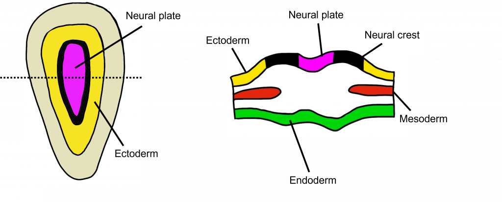
Very early in development, an embryo looks like a flat disc with 3 different layers of cells.
- Endoderm: will eventually develop into the viscera (organs of the body)
- Mesoderm: will eventually develop into the bones and muscles
- Ectoderm: will eventually develop in the nervous system and the skin
The Neural Tube
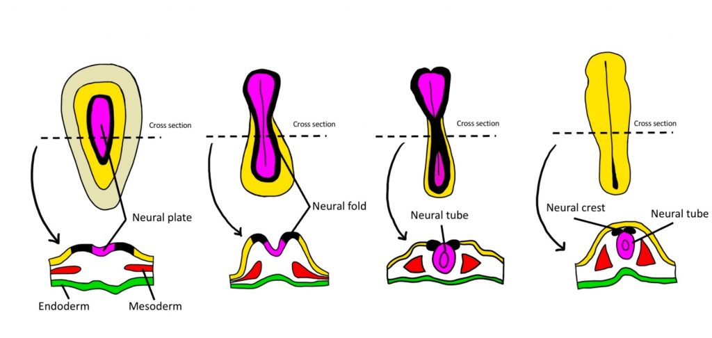
Let’s focus on the ectoderm that will develop into the nervous system. The ectoderm layer will fold together in a process called neurulation to form a neural tube. Neurulation occurs within the first month following conception in humans.
During neurulation, the middle of the neural plate folds first, followed by the anterior and posterior ends. The central nervous system will develop from the walls of the neural tube and the hollow lumen of the tube will become the cerebral ventricles and spinal canal within the central nervous system.
Neural Tube Defects
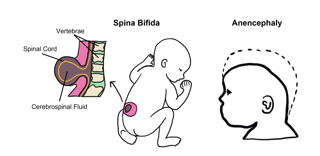
Early in pregnancy, there can be problems with the process of neurulation and the formation and closure of the neural tube. Collectively, these are referred to as neural tube defects that include defects that occur within the brain, spine, or spinal cord. One of the more common neural tube defects is spina bifida. In spina bifida, the neural tube fails to close completely, and as a result the backbone that typically protects the spinal cord also does not form appropriately. This event can occur anywhere along the spine, and will typically result in nerve damage at that particular site that will potentially lead to physical and / or intellectual disability ranging in severity with the degree of damage.
Anencephaly is a neural tube defect that is caused by the failure of the anterior portion of the neural tube to close. Anencephaly is a serious birth defect that typically prevents the development of the anterior portion of the brain and associated skull.
Unfortunately, many neural tube defects occur very early in pregnancy, such that many women do not yet know that they are pregnant. As a result, daily folic acid supplements or a diet that includes foods rich in folate is recommended to all women of reproductive age to help prevent neural tube defects in the event of a pregnancy.
Three Vesicle Stage of Development
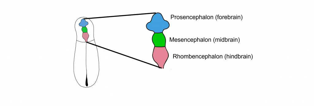
Early in development, the anterior portion of the neural tube has three distinct vesicles, which will each develop into different structures. These vesicles, from most anterior to most posterior, are the prosencephalon (forebrain), the mesencephalon (midbrain), and the rhombencephalon (hindbrain).
Through development, the walls of these vesicles will differentiate into adult brain structures. Differentiation is the process by which structures become more complex and functionally specialized during development. The names of these early vesicles can be used to describe either the stages through development, or a grouping of structures that eventually form in adulthood. In the following chapters, we will learn more about some of these anatomical structures.
Prosencephalon (Forebrain)
The prosencephalon, or the forebrain, is most anterior in the neural tube and eventually develops into the “higher order” brain regions, including the cerebral cortex. Most of the time, when you see an image of an intact brain from the side or the top, the structures that are visible to you are the forebrain structures. The prosencephalon is made up of the telencephalon and the diencephalon.
As the prosencephalon continues to develop, additional structures will form that are associated with the cerebral hemispheres. For instance, the optic vesicles that bud off the surface of the prosencephalon will differentiate into the retinas of the eyes—thus, the retina is made up of neural tissue. The olfactory bulbs will differentiate from the ventral surface of the cerebral hemispheres.
Note that as the walls of the tube develop into these structures, the lumen of the tube remains and will become the fluid-filled ventricles of the brain. The lateral ventricles will form from the tube within the cerebral hemispheres and the third ventricle will be found ventrally surrounded by the diencephalon.
Structures of the Telencephalon: Cerebral Cortex and the Basal Ganglia
The cerebral cortex makes up the outermost layer of the brain. The word “cortex” comes from the word meaning “bark”, the outer layer of a tree. The cerebral cortex is the most evolved structure of the human brain and is responsible for higher order thinking. Here, the brain processes behaviors such as attention, memory, and language.
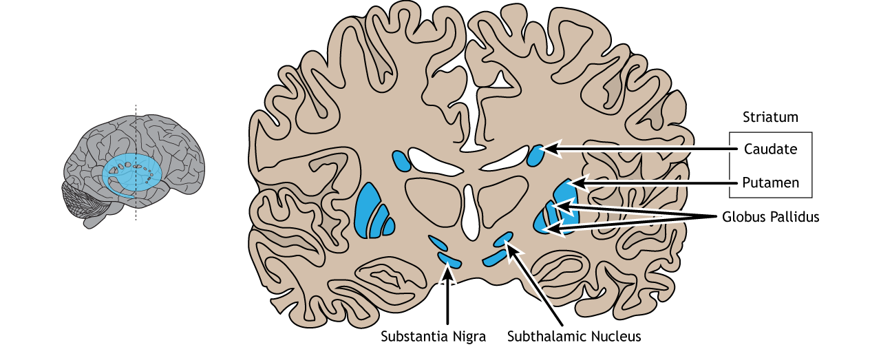
The basal ganglia are made up of a series of brain structures (including the caudate and putamen, globus pallidus, substantia nigra, and subthalamic nucleus) that are used for such behaviors as motor and habit learning, emotional processing, and action selection.
Structures of the Diencephalon: Thalamus and Hypothalamus
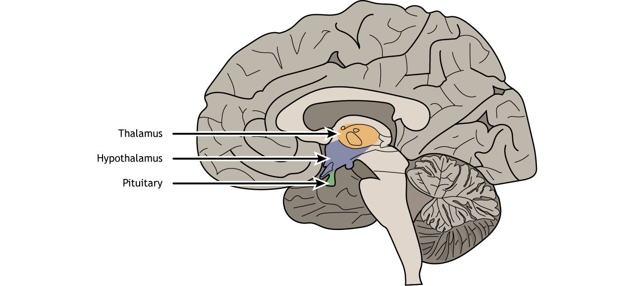
The thalamus is often referred to as a “sensory relay station” in the brain, since almost every sensory modality (sight, taste, touch, and hearing) passes information through the thalamus before being directed to the appropriate area of the cortex.
The hypothalamus is also within the diencephalon. This structure serves as an autonomic control center to alter visceral function and a communication route to the body’s endocrine system through control of anterior pituitary hormones and production of posterior pituitary hormones. Neural signals originating in the hypothalamus have the capability to influence the chemistry and function of the entire body. The hypothalamus also has nuclei that function in emotional responses, and regulate body temperature, food intake, water balance, and sleep.
Mesencephalon
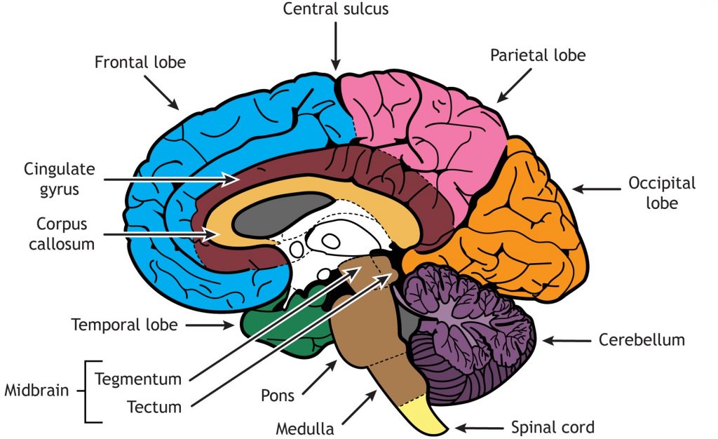
Moving more caudally, the mesencephalon (or the midbrain) differentiates into the tectum and the tegmentum.
Tectum
The tectum (which means “roof”) is the dorsal portion of the midbrain and it has two major structures: the superior colliculus and inferior colliculus. The superior colliculus is important in reflexive eye movements that allow you to quickly orient to something changing in your environment. The inferior colliculus relays auditory information to the thalamus for auditory function.
Tegmentum
The tegmentum (which means “floor”) is the ventral portion of the midbrain. There are many structures in the tegmentum and they can perform a wide variety of functions. For example, the periaqueductal gray allows us to respond to painful stimuli, the red nucleus and substantia nigra coordinate complex movements, and the ventral tegmental area is important for the processing of reward and motivation.
Rhombencephalon
The most posterior/caudal portion of the neural tube is the rhombencephalon (or hindbrain). Evolutionarily speaking, the rhombencephalon represents the oldest part of the central nervous system. These structures likely evolved some 570 million years ago.
Rostral Hindbrain
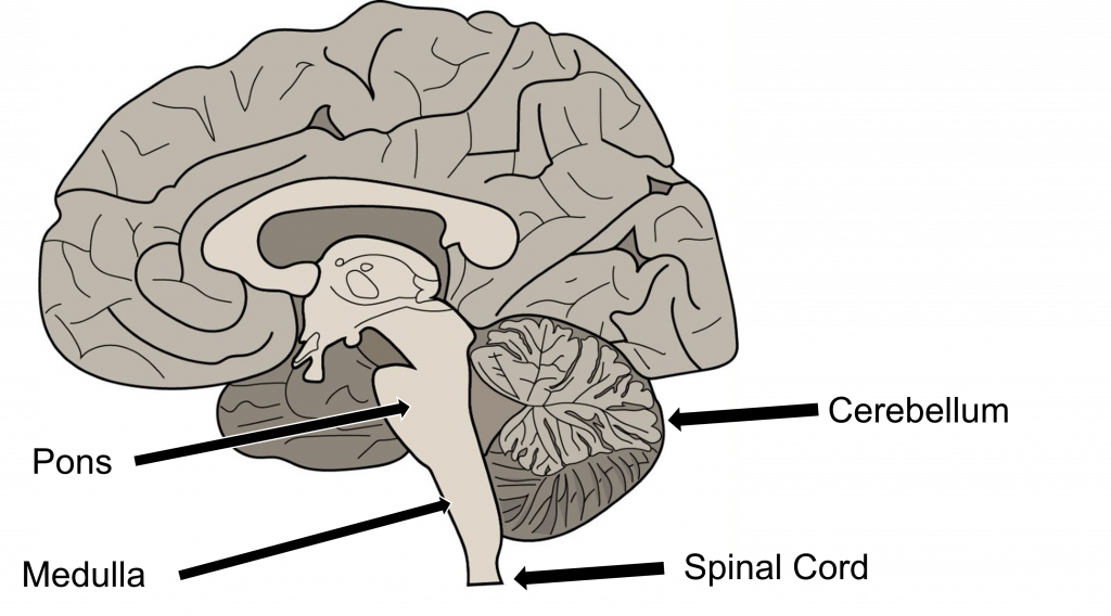
The more rostral portion of the hindbrain differentiates into two structures: the cerebellum and the pons. The cerebellum, or “little brain”, is best known as a structure that enables motor control functions, such as balance, coordination, posture, and learning physical actions. The cerebellum helps us recognize and predict sequences of events during motor learning. More recently, the cerebellum has also been recognized to play a role in non-motor functions like learning. The pons (which means “bridge”) is an important structure that relays impulses between the motor cortex and the cerebellum. It has a critical function to help us perform involuntary functions like breathing.
Caudal Hindbrain
The caudal portion of the hindbrain differentiates into the medulla. The medulla is found at the far posterior end of these three early developmental vesicles. The medulla contains many clumps of neurons that are responsible for functions that an organism carries out unconsciously. It contains a cardiovascular center that is especially important in maintaining and changing blood pressure and heart rate, and also has connections to the pons to help regulate breathing. The medulla contains areas that can detect toxins in the blood that come from dietary sources, triggering vomiting. Other behaviors like hiccupping, swallowing, coughing, and sneezing are also controlled by the medulla.
Moving beyond this anterior portion of the neural tube that contains the prosencephalon, mesencephalon, and rhombencephalon, the neural tube continues down the length of the embryo. The remainder of the neural tube will differentiate into the spinal cord, with the lumen of the tube becoming the central canal of the spinal cord. The spinal cord is important in relaying information to and from the body.
Key Takeaways
- The ectoderm layer of the embryo develops into the nervous system
- Neurulation is the process that forms the neural tube and failure for portions of the neural tube to close can cause neural tube defects
- The prosencephalon, mesencephalon, and rhombencephalon are the three vesicles at the anterior end of the neural tube. They will develop into structures in the adult brain.
Test Yourself!
Attributions
Portions of this chapter were remixed and revised from the following sources:
- Foundations of Neuroscience by Casey Henley. The original work is licensed under a Creative Commons Attribution-NonCommercial-ShareAlike 4.0 International License
- Open Neuroscience Initiative by Austin Lim. The original work is licensed under a Creative Commons Attribution-NonCommercial 4.0 International License.
Media Attributions
- Three layers of the embryo © Valerie Hedges is licensed under a CC BY-NC-SA (Attribution NonCommercial ShareAlike) license
- Neurulation © Valerie Hedges is licensed under a CC BY-NC-SA (Attribution NonCommercial ShareAlike) license
- Neural Tube Defects © Valerie Hedges is licensed under a CC BY-NC-SA (Attribution NonCommercial ShareAlike) license
- 3 vesicle stage of development © Valerie Hedges is licensed under a CC BY-NC-SA (Attribution NonCommercial ShareAlike) license
- Sagittal section of brain structures © Casey Henley is licensed under a CC BY-NC-SA (Attribution NonCommercial ShareAlike) license
- Structures of the rhombencephalon © Valerie Hedges is licensed under a CC BY-NC-SA (Attribution NonCommercial ShareAlike) license
Folding of the ectoderm that results in the structure of the neural tube
Cavities within the brain that are filled with cerebral spinal fluid.
Birth defects caused by failure of the neural tube to close completely
in front of; toward the face
Most posterior portion of the neural tube. Also called the hindbrain.
Most posterior portion of the neural tube. Also called the rhombencephalon.
