
4 Visualizing Cells of the Nervous System
For the majority of human history, the only way we were able to study the structure of the brain was with crude, butcher-like methods. Wait for a person to die, saw a giant hole in the top of the skull, take the brain out, and slice it into pieces to see if there was some correlation between the way the brain looks and the way they died. With these methods, only major changes in gross anatomy could be observed, such as those resulting from severe birth defects or trauma.
Brain analysis methods were enhanced by the scientific adoption of light microscopy. Naturalists in the mid 1600s such as Antonie van Leeuwenhoek, Jan Swammerdam, and Robert Hooke began looking at biological substances up close, and the brain proved to be a complex and interesting sample of tissue.
Visualizing Cells: Cell Staining
Staining is an imaging method that is often used in conjunction with microscopy. Thin slices of brain tissue are exposed to various chemical processes. The chemicals that are used for staining have different affinities for parts of cells.
For staining to work, the tissue needs to be subjected to a series of chemical processes. First, the tissue needs to be fixed. Fixation is a chemical process that is accomplished by exposing the tissue to a chemical like paraformaldehyde. The most effective way to expose every part of the body to fixative is to “hijack” the endogenous circulatory system by flushing fixative through the arteries, a process called perfusion. Chemically, fixatives cause adjacent proteins to become covalently bonded (crosslinked), a process which causes the proteins to become unchangeable; they become “fixed” in time. These fixatives are very harsh chemicals, and usually kill microorganisms and inactivate the endogenous enzymes that normally degrade biological tissue. (As a side note, fixatives are particularly nasty carcinogens that can permeate easily through latex gloves.)
After fixation, devices such as a microtome or cryostat are used to slice the brain into sections as thin as 10 microns. Stains do not always reliably pass all the way through thick sections of tissue, such as an intact brain. With thin slices, chemical stains are able to permeate through the depth of the tissue.
Next, stains are used to color the cells of the nervous system. This process is used because it is good for identifying the location of specific proteins at a subcellular level.
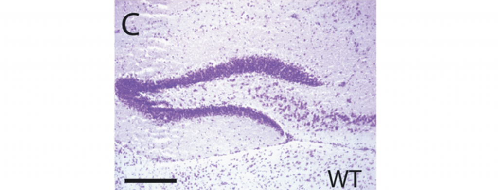
Nissl-Staining (Cresyl Violet Staining)
A stain is needed to distinguish individual cells in nervous tissue.
Nissl stain (Cresyl Violet Stain) was discovered by Franz Nissl. It stains nucleic acids such as RNA and DNA. Thus, the stain is only localized to neuronal cell bodies, where RNA and DNA are found. This staining method is useful for studying neuronal arrangement and how densely neurons are packed in specific brain structures.
Golgi Staining
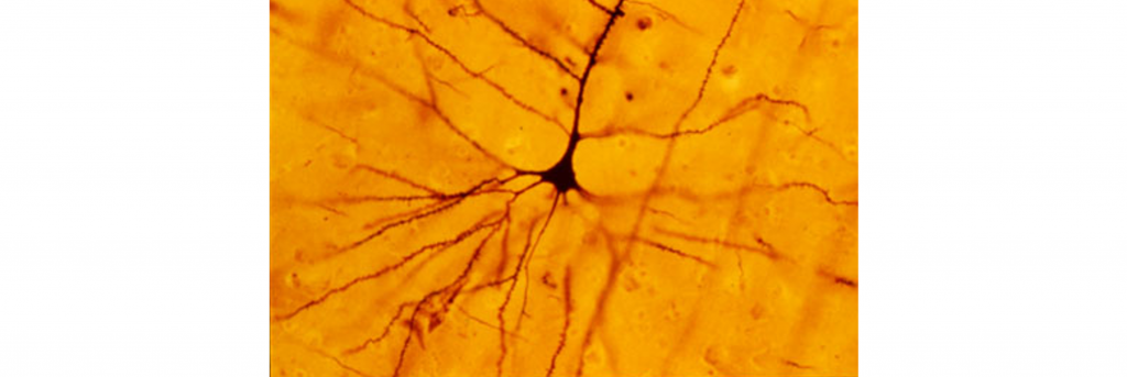
A major advancement in the study of neuronal morphology came about in the late 1800s. The Italian anatomist and biologist Camillo Golgi identified a shortcoming with the cellular analysis techniques of the time: structures in the central nervous sytem were impossible to distinguish from one another. The cells in the brain were so densely packed together, that it became difficult to identify which cellular material belonged to which cell.

Golgi came up with a new technique using a silver compound that caused the silver to precipitate inside the cell membranes. However, not every cell took up the silver. Instead, only a small fraction of neurons, maybe 1% or even less, were completely stained in black, which stood out remarkably well against the light yellow background of the surrounding tissue. This reaction, initially called the “black reaction”, is now known as a “Golgi stain”. (Despite being more than a hundred years old, we currently don’t know the mechanism by which the silver stain is taken up into the neurons, or what determines why certain cells take the stain and others don’t.)
Because of the great contrast between cell and background, every single part of the neuron was completely filled, allowing Golgi to do drawings of the morphology of this nervous tissue. Based on his staining results, Golgi supported the idea that the parts of the nervous system are all one very large, physically connected network. This idea was known as the Reticular Theory.
Santiago Ramon y Cajal
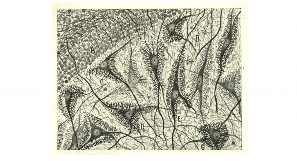
About 10 years later, the Spanish neuroanatomist Santiago Ramon y Cajal repeated some of Golgi’s staining experiments with other sections of nervous tissue. Looking at similar darkly-filled neurons, Cajal arrived at a different conclusion: the nervous system is not a giant net, but rather a series of individual units that are separated from one another physically. This idea came to be known as the Neuron Doctrine.
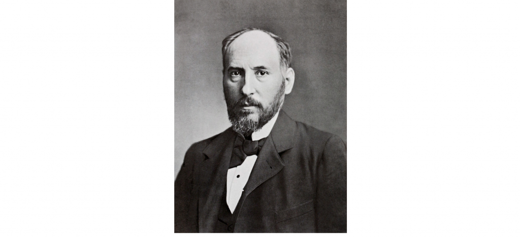
Both Golgi and Cajal were awarded the shared Nobel Prize in Physiology or Medicine in 1906 for their accomplishments in helping to understand “the structure of the nervous system”. Even though Cajal’s Neuron Doctrine was adopted widely by scientists, the elucidation of this organization would not have been made possible without Golgi’s development of the silver stain. The sharing of this prestigious award was ironic because of the many disagreements between the two scientists.
Cajal’s Neuron Doctrine was eventually given more support with the aid of modern techniques, like electron microscopy, that are capable of physically seeing the distance between two neurons. The Neuron Doctrine represents our current understanding of how the nervous system is organized.
Key Takeaways
- Nissl stains stain neuronal cell bodies
- Golgi stains stain entire neurons and allows for visualization of cell structures (dendrites and axons)
- Neurons are discreet cells and are not interconnected together together into a network
Test Yourself!
Attributions
Portions of this chapter were remixed and revised from the following sources:
- Open Neuroscience Initiative by Austin Lim. The original work is licensed under a Creative Commons Attribution-NonCommercial 4.0 International License.
Media Attributions
- Nissl Stain © Marika Nosten-Bertrand, Caroline Kappeler, Céline Dinocourt, Cécile Denis, Johanne Germain, Françoise Phan Dinh Tuy, Soraya Verstraeten, Chantal Alvarez, Christine Métin, Jamel Chelly, Bruno Giros, Richard Miles, Antoine Depaulis et Fiona Francis adapted by Valerie Hedges is licensed under a CC BY (Attribution) license
- Camillo Golgi © Unknown adapted by Valerie Hedges is licensed under a CC BY (Attribution) license
- Golgi Stain © Cahass adapted by Valerie Hedges is licensed under a CC BY-SA (Attribution ShareAlike) license
- Santiago Ramon y Cajal © Anonymous adapted by Valerie Hedges is licensed under a Public Domain license
- Santiago Ramon y Cajal drawing © Santiago Ramon y Cajal adapted by Valerie Hedges is licensed under a Public Domain license
Theory that all neurons are interconnected in one giant network
The idea that neurons are not interconnected but rather exist as discreet units
