
18 Neurotransmitters: Catecholamines (Dopamine, Norepinephrine, Epinephrine)
Catecholamines are a class of neurotransmitters that are found within the larger class of neurotransmitters, biogenic amines. The catecholamines share many characteristics.
Dopamine
Dopamine, a catecholamine transmitter, plays many roles in the nervous system, but it is best known for its roles in reward and movement.
Synthesis
In the presynaptic terminal, the amino acid tyrosine is converted into DOPA via tyrosine hydroxylase, which is the rate-limiting step in the synthesis of all the catecholamines. DOPA is then converted to dopamine by DOPA decarboxylase. Dopamine is packaged into small synaptic vesicles by the vesicular monoamine transporter (VMAT).
Unlike glutamate or GABA, dopamine-producing neurons are not widely abundant in the brain. Instead, there are generally only a few patches of neurons that produce dopamine, most of which are found in the midbrain. Two areas include the ventral tegmental area and the substantia nigra.
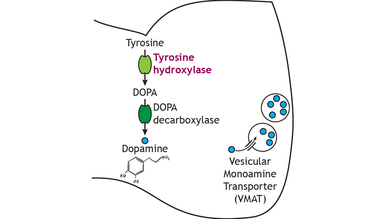 Figure 18.1. Dopamine is synthesized in a two-step process. Tyrosine is converted into DOPA by tyrosine hydroxylase, the rate-limiting step in the pathway. Then dopamine is synthesized from DOPA by DOPA decarboxylase. Dopamine is then packaged into vesicles by vesicular monoamine transporter. ‘Dopamine Synthesis’ by Casey Henley is licensed under a Creative Commons Attribution Non-Commercial Share-Alike (CC BY-NC-SA) 4.0 International License.
Dopamine Receptors
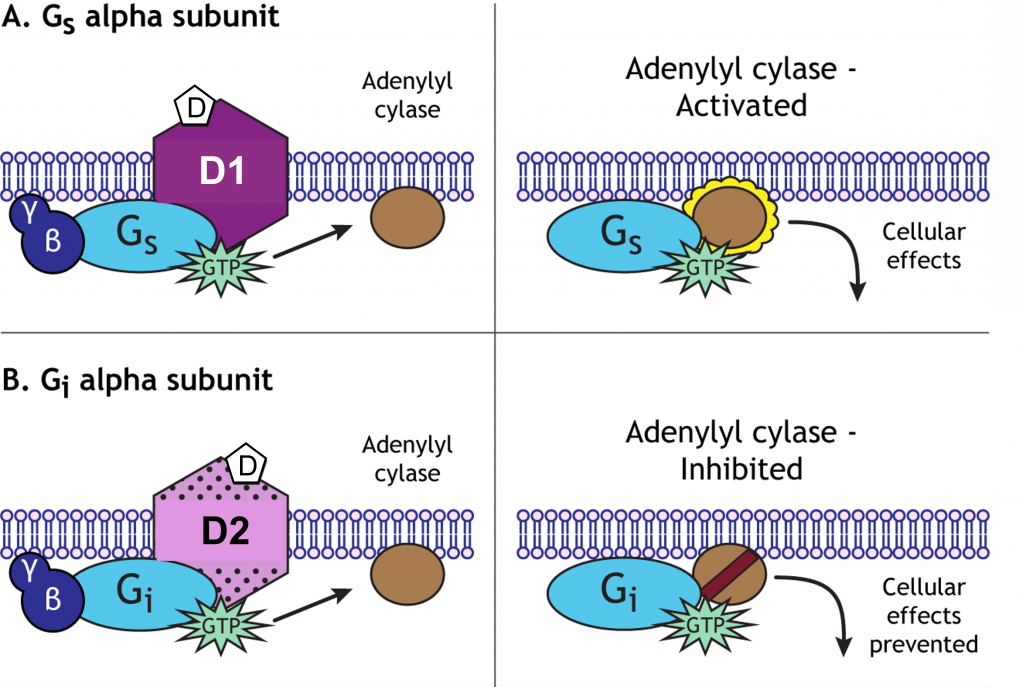
Dopamine binds to metabotropic receptors on postsynaptic cells. There are two classes of dopamine metabotropic receptors: D1-like receptors and D2-like receptors. D1-like receptors are typically associated with Gαs and cause activation of the cAMP/adenylyl cyclase second messenger system and the generation of EPSPs in the postsynaptic cell.
D2-like receptors, however, are typically associated with Gαi that inhibits the cAMP/adenylyl cyclase second messenger system and generate IPSPs in the postsynaptic cell.
Termination of Dopamine Signaling
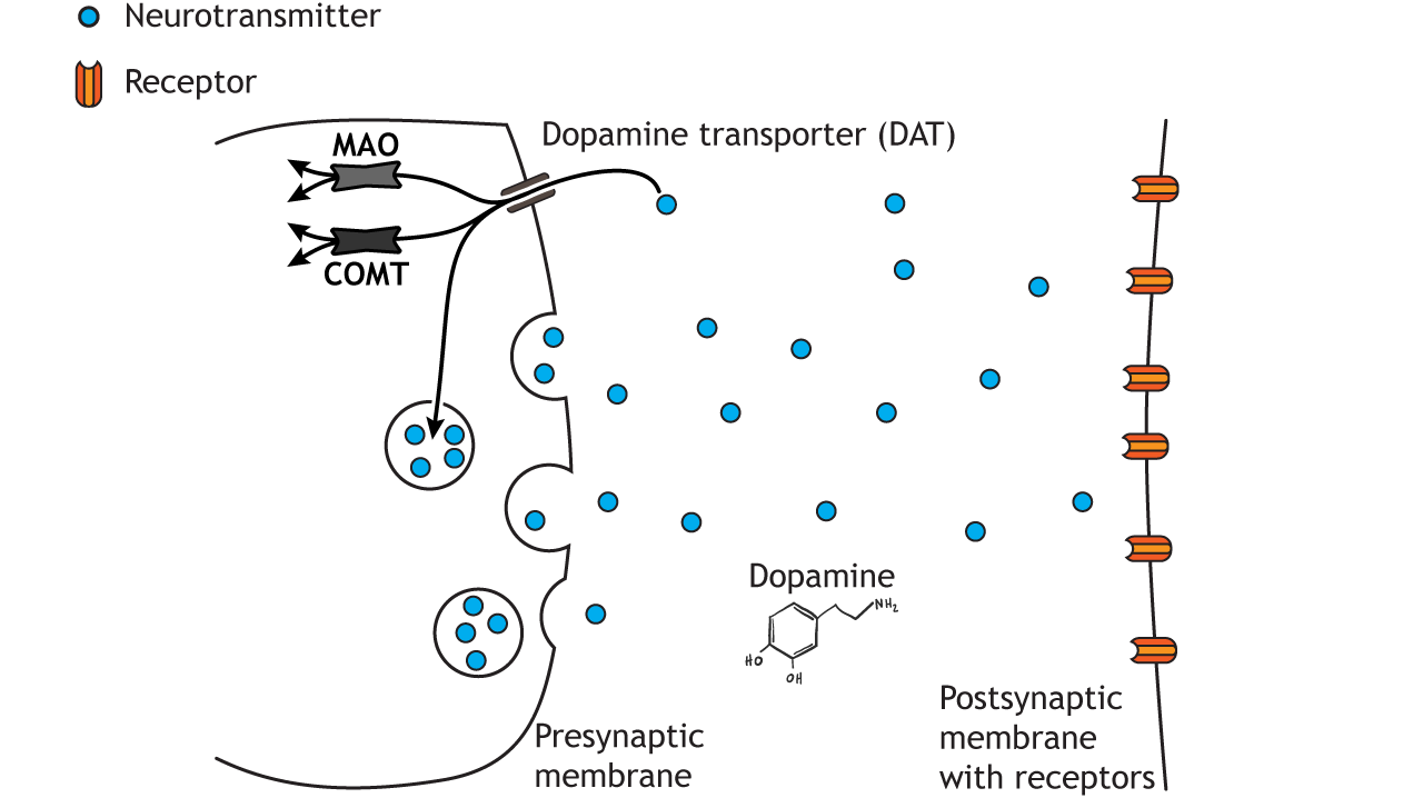
Dopamine action is terminated by reuptake into the presynaptic terminal via the dopamine transporter (DAT). Once inside the cell, dopamine is either degraded via the actions of either monoamine oxidase (MAO) or catechol-O-methyltransferase (COMT), or it is repackaged into vesicles.
Norepinephrine
Norepinephrine-producing cells are localized in the pons of the brain stem, a structure called the locus coeruleus. The locus coeruleus is very small, but these neurons send projections widely throughout the brain. Outside of the brain, we think of norepinephrine as being responsible for triggering the sympathetic nervous system response of the body, the “fight-or-flight” reaction that prepares the body for physical activity in times of fear or acute stress.
Synthesis
In neurons that release norepinephrine, which is another catecholamine transmitter, once dopamine is packaged into small synaptic vesicles, a membrane-bound enzyme called dopamine beta-hydroxylase converts dopamine into norepinephrine. Therefore, unlike the other small molecule neurotransmitters, norepinephrine is synthesized within the vesicles, not in the cytoplasm. Like dopamine, the rate-limiting step of this synthesis pathway is the activity of tyrosine hydroxylase.
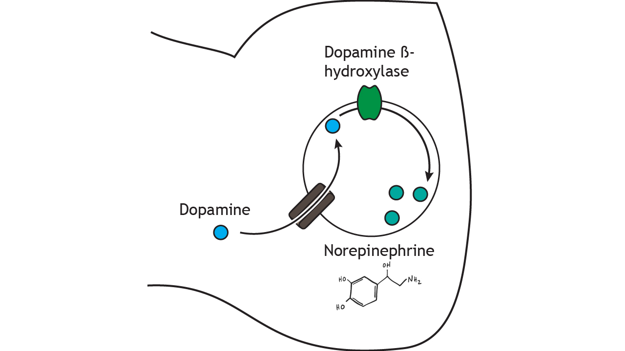 Figure 18.4. Norepinephrine is synthesized from dopamine by dopamine beta-hydroxylase after packaging into vesicles. ‘Norepinephrine Synthesis’ by Casey Henley is licensed under a Creative Commons Attribution Non-Commercial Share-Alike (CC BY-NC-SA) 4.0 International License.
Norepinephrine Receptors
Norepinephrine binds to metabotropic adrenergic receptors (α1, α2, and β). The binding of norepinephrine to its receptor activates second messenger signaling cascades that will cause either EPSPs or IPSPs, depending on the receptor subtype.
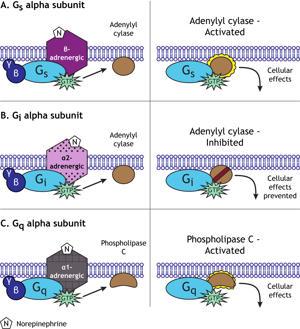 Figure 18.5. The norepinephrine beta-adrenergic receptor couples to the Gαs subunit and activates adenylyl cyclase, which initiates downstream cellular effects. B) The norepinephrine alpha 2-adrenergic receptor couples to the Gαi subunit and inhibits adenylyl cyclase, which prevents downstream cellular effects. C) The norepinephrine alpha 1-adrenergic receptor couples to the Gαq subunit and activates phospholipase C, which initiates downstream cellular effects. ‘Alpha Subunit Effects’ by Casey Henley is licensed under a Creative Commons Attribution Non-Commercial Share-Alike (CC BY-NC-SA) 4.0 International License.
Termination of Norepinephrine Signaling
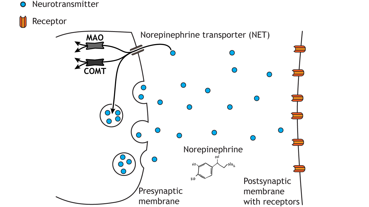
Termination of norepinephrine signaling is similar to the termination of dopamine signaling. Reuptake into the presynaptic terminal occurs via the norepinephrine transporter (NET), and then the transmitter is either degraded within the cell by MAO or COMT or repackaged into synaptic vesicles.
Epinephrine
Epinephrine, also called adrenaline, is a catecholamine, but it is often considered a hormone instead of a neurotransmitter. Epinephrine is primarily released by the adrenal medulla into the circulation; it is used as a neurotransmitter in only a small number of neurons.
Synthesis
Epinephrine is synthesized from norepinephrine in the cytoplasm by the enzyme phenylethanolamine-N-methyltransferase, so epinephrine synthesis requires norepinephrine to exit the vesicles where it was synthesized. After synthesis in the cytoplasm, epinephrine is repackaged into small vesicles via the vesicular monoamine transporter.
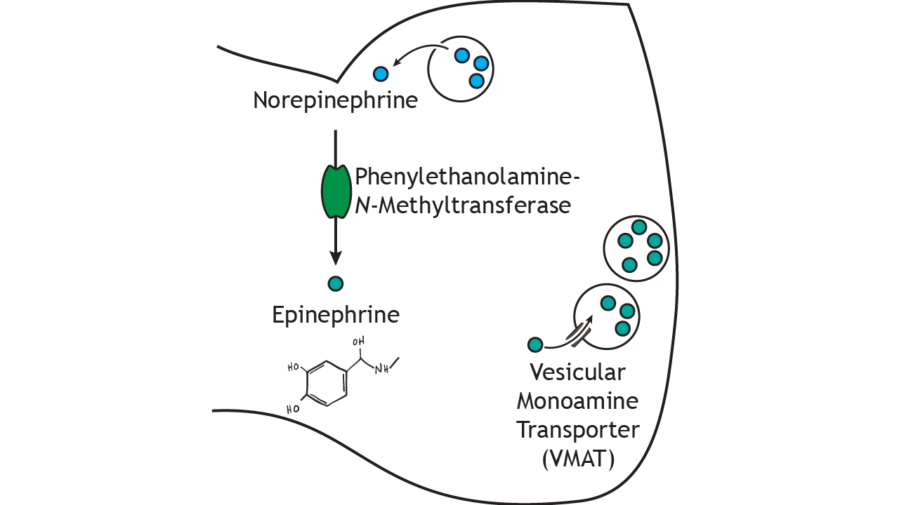 Figure 18.7. Epinephrine is synthesized from norepinephrine by phenylethanolamine-N-methyltransferase in the cytoplasm. Epinephrine is then packaged into vesicles by vesicular monoamine transporter. ‘Epinephrine Synthesis’ by Casey Henley is licensed under a Creative Commons Attribution Non-Commercial Share-Alike (CC BY-NC-SA) 4.0 International License.
Epinephrine Receptors
Epinephrine also binds to α and β adrenergic receptors (described above for norepinephrine) and causes similar activity when bound to these receptors.
Termination of Epinephrine Signaling
Epinephrine, similarly to norepinephrine, also goes through reuptake into the presynaptic cell. Further, it is also degraded within the cell by both MAO or COMT or repackaged into synaptic vesicles.
Key Takeaways
- The catecholamines are synthesized from the amino acid tyrosine
- The synthesis pathways overlap and share many enzymes and proteins
- The catecholamines bind to metabotropic receptors
- The catecholamines are inactivated via similar mechanisms: reuptake, MAO and COMT
Test Yourself!
Attributions
Portions of this chapter were remixed and revised from the following sources:
- Foundations of Neuroscience by Casey Henley. The original work is licensed under a Creative Commons Attribution-NonCommercial-ShareAlike 4.0 International License
- Open Neuroscience Initiative by Austin Lim. The original work is licensed under a Creative Commons Attribution-NonCommercial 4.0 International License.
Media Attributions
- Dopamine Receptors © Casey Henley adapted by Valerie Hedges is licensed under a CC BY-NC-SA (Attribution NonCommercial ShareAlike) license
Process where neurotransmitters are removed from the synapse and transported back into the presynaptic cell to be re-packaged into synaptic vesicles
Postsynaptic receptors in which neurotransmitters bind and cause the activation of an associated G-protein and cell signaling cascades. The affected ion channel and receptor are found on different transmembrane proteins..
