
1 What is Neuroscience?
Learning Objectives
- Define neuroscience and explain its role in understanding the nervous system.
- Identify the major disciplines that contribute to the field of neuroscience and explain their unique perspectives, recognizing the interdisciplinary nature of neuroscience.
- Explain the importance of experimental design in neuroscience research, including the roles of independent and dependent variables, control groups, and confounding variables.
- Discuss the significance of case studies in neuroscience, such as the key historical examples of Phineas Gage and Patient HM.
- Compare and contrast different research methods used to study the nervous system.
What is Neuroscience?
Neuroscience is the study of the nervous system, the collection of nerve cells that interpret all sorts of information which allows the body to coordinate activity in response to the environment.
The study of neuroscience has taught us that the brain is a complicated organ with several connection routes, both between different bodily organs and within itself. Some of those connections communicate information down towards the body, such as signals that allow us to control the movements of our muscles or to change the activity of our internal organs. Other connections ascend into the brain, conveying all sorts of information from the world around us into a representation of our surroundings. Still, other routes communicate between brain areas, such as when the sudden detection of a threat passes through our visual system and turns into a “get ready” signal that then prepares the rest of our body for conflict. Because of this complex system of communication, the nervous system can be thought of as a series of highways and roads that connect different cities (organs).
The nervous system conveys all of these different types of information using a combination of electrical and chemical signals. The main active cellular units of the nervous system, the neurons, are highly sensitive to changes in their environment. A wide variety of chemicals called neurotransmitters are responsible for passing information between neurons.
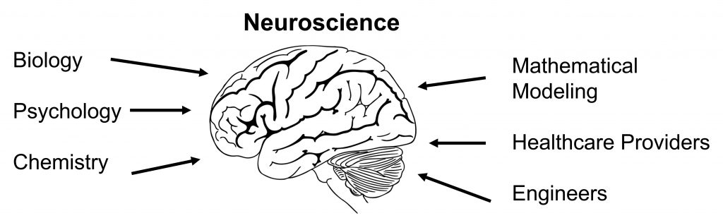
Neuroscience is an integrative field of study
Realistically, our modern understanding of “neuroscience” is a combination of several academic disciplines, all using their strengths to understand some aspect of the nervous system. Because of this integrative nature, it is possible to study neuroscience from many different perspectives, each of them more fitting for answering different types of questions. These “angles” of analysis are described below.
Biology
At the root of the study is biology. Whenever you are studying living processes, such as learning, visual perception, or consciousness, you dip into the realm of biology. The broad field of biology can be subdivided into smaller, more precise categories. Molecular neurobiologists study proteins and gene regulation, cellular neurobiologists examine how networks of neurons communicate with one another, and cognitive neuroscientists study the underlying causes of behaviors. Understanding neuroscience involves genetics, such as the autosomal dominant neurodegenerative condition Huntington’s disease. Other biological sub-disciplines, such as ecology and evolution, are also considered in neuroscience as well, such as the parasite Toxoplasma, which changes an animal’s response to fearful stimuli, allowing the organism to reproduce as it moves through different species in the food web.
Psychology
Psychology provided the earliest explanations about the brain and ideas about the origin of the mind. Some questions in this field branched off from philosophy as people began thinking about the “mind–body problem”, the discussion that centered around the question of whether a function as complex as consciousness could result from the activity of a clump of cells. Psychologists also wondered whether parts of the brain in isolation have different properties than when those parts are working together. This property, called emergence, is the idea that the whole is greater than the sum of its parts. Psychologists examine neuroscience from a top-down view, aiming questions at understanding the whole organism before looking at smaller components of the organism (compare this with biological approaches, often a bottom-up view that starts at the level of cells or molecules).
Chemistry
Chemistry is a strong influencer of nervous system function—just ask anyone who forgot their morning cup of coffee! We use a variety of endogenous (originating from within the body) chemicals that act as signaling molecules, allowing communication between cells. These chemicals exist in many different structures, which determine their function; some are acidic while others are basic, some are polar, others are fat-soluble, and some are even gases. The nervous system is also highly sensitive to influence by exogenous chemicals (meaning they originate from outside the body), such as caffeine and cocaine.
Physics
Many principles of physics can be observed through the functioning of neurons. For example, neurons maintain a negative electrical charge, usually measured on the scale of tens of millivolts (a millivolt is a thousandth of a volt.) The main way for neurons to send signals depends on a temporary change in this voltage; this signal is called an action potential. This change in voltage is brought on by the movement of charged ions across the cell membrane, and they closely follow the rules of magnetism: opposite charges attract while like charges repel.
Mathematical Modeling
The field of computational neuroscience has grown from the use of mathematical modeling to describe or predict some aspect of the nervous system. If our current estimates are correct, we have around 86 billion neurons in the brain, a number so large that it is difficult to conceptualize. It would be nearly impossible to understand that many components of a system without taking advantage of the sheer mathematical strength of a computer.
Healthcare Providers
Healthcare providers, like neurologists and psychiatrists, work from a different angle. They coordinate closely with researchers to apply scientific knowledge from the field or laboratory to treat patients, thus using biological principles as therapies. For example, neurologist Dr. Oliver Sacks used his knowledge of the dopamine neurotransmitter system to treat patients with a paralysis-like condition in the 1960s, leading to the development of levadopa treatment for Parkinson’s disease. Other healthcare providers use imaging strategies like a CT scan to assess the extent of a head injury or the location of a brain tumor, while an EEG can be helpful for the diagnosis of epilepsy.
Engineers
Engineers help develop the tools needed to understand questions in neuroscience, such as the patch clamp rig or electron microscope, highly specialized pieces of lab equipment. They also work closely with healthcare providers to translate science into therapy, such as the deep brain stimulator devices for the treatment of conditions such as Parkinson’s disease. Collectively, all the people who participate in neuroscience in some way are united by their interest in the workings of the body. Because of the overwhelming complexity of the nervous system, there are many questions still unanswered. The continual appearance of new questions in neuroscience keeps us wondering, inspires curiosity, and promises a multitude of fascinating career paths for centuries to come.
How do we learn about neuroscience?
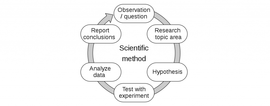
Experimental design
The gold standard in science is the use of experimental design. In an experiment, the scientist uses a stepwise process of developing a research question and hypothesis, then answering that question by performing tests. The main goal of an experiment is to establish a causal relationship between one factor that is being changed, the independent variable, and the factor that is influenced, the dependent variable. A well-designed experiment has variables that are carefully controlled, which minimizes the influence of extraneous variables, often called confounding variables. The influence of confounding variables can be eliminated by comparing the experimental group with a control group, a group that is as similar as possible in every way except for the manipulation of the independent variable. Importantly, subjects or patients are generally assigned to the experimental or control group at random.
 Case Studies
Another strategy is the case study, a highly detailed description of a single patient and their condition. A case study documents the details regarding a specific deficit or enhancement and is an opportunity to examine individuals with very rare conditions, which are useful for informing about the functions of different brain structures. Like a quasi-experimental study, case studies only show correlation, not causation. It is difficult to generalize the findings from a case study to the population at large.
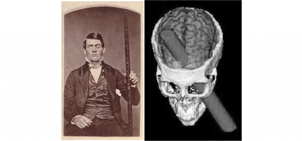
Perhaps the most famous case study in all of neuroscience is the 1848 story of the railroad worker Phineas Gage. Gage was a construction foreman working on the railroad when an unfortunate explosive workplace accident caused a iron rod to be driven through his left frontal lobe, largely destroying it. Remarkably, Gage survived this accident and lived another 12 years. However, Gage’s acquaintances described subsequent changes in his personality, teaching us that one of the functions of this area of the brain is regulating our inhibitions.
Case studies can be helpful for the development of hypotheses that can later be tested experimentally. For example, consider another famous case study of Patient HM, the man who had his left and right hippocampus surgically removed and couldn’t create certain types of memory. A research question based on this case study might be: “Is the hippocampus needed for the creation of navigational memory?” Then, an experimental study could be performed in rodents, where we surgically remove the hippocampus (experimental group) or a different part of the brain (control group) and see how well the rodents perform on a memory task.
The Use of Animals in Research
Though there are many ways that we can directly study humans through experimentation or case studies, it is often impossible to test every question in humans. Instead of always studying humans, scientists often use nonhuman model organisms, the most common organisms being the worm C. elegans, fruit flies (Drosophila melanogaster), zebrafish (Danio rerio), song birds, mice, rats, and macaque monkeys.

The closer we move towards the human, the more similarities the model organism shares with us. Of the commonly used model organisms, macaque monkeys are the non-humans that are most similar to humans. We share 93% of our genetic material with macaques, but we still have different metabolic and physiological processes, and our behaviors are much different from theirs. Ethical constraints prevent us from performing experiments that may cause physical or psychological harm if performed in humans. We would never conduct a test on humans to assess what concentration of neurotoxin leads to brain damage (these experiments aren’t done very frequently in nonhumans anyway). Invertebrates, such as worms and fruit flies are not as heavily regulated by ethics oversight committees, allowing scientists to conduct a wider set of experiments on these animals.
Our moral responsibilities toward animal subjects are that:
- Animals should only be used in worthwhile experiments.
- All steps are taken to minimize pain and distress.
- All possible alternatives to animal research are considered.
Research facilities at colleges and universities are monitored by an Institutional Care and Use Committee (IACUC). The IACUC consists of fulltime veterinarians, scientists, and community members. They must follow federal laws when approving animal research.
Experimental Preparations
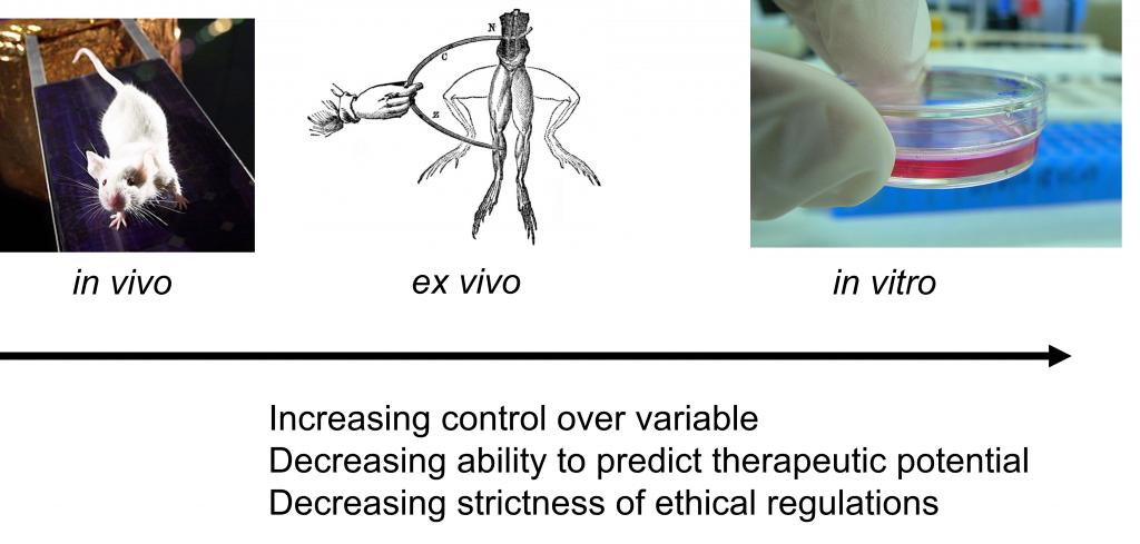
Performing an experiment in an intact, living organism, whether human or nonhuman, is described as an in vivo (Latin meaning “within life”) preparation. The main strength of this strategy is that the data collected here are more predictive of the human condition, which is one of the main goals of biomedical research. However, the in vivo preparation has challenges, because thousands of variables within a living system are uncontrolled or still unknown. There are also very strict ethical limitations on the nature of experiments that can be done in vivo.
On the other hand, an in vitro (Latin meaning “within glass”) preparation is an experiment performed on cultured cells or isolated molecules of DNA, RNA, or protein. These preparations have the opposite strengths and weaknesses of in vivo preparations. They allow for extremely good control over variables, but the results are less reliable in translating to a therapy. The regulations on these experiments are much more lax compared to in vivo experiments; most of the regulatory guidelines are to protect the experimenter rather than the patient or the experimental subject.
Falling in between these two preparations is an ex vivo experiment. In this kind of experiment, a section of the living organism is taken, such as a slice of brain, a tissue biopsy, or a detached frog leg. The strengths and limitations of these experiments are somewhere in between that of the other two preparations.
What neuroscience is not
As complex as the brain is, naturally misconceptions make their way into popular culture. It’s valuable to address these myths about neuroscience and explain the evidence that refutes these statements .
Myth 1: “We only use 10% of our brain.”
This wildly inaccurate statistic has been the foundation for several fictional movies, TV shows, and books. The truth is that we use every part of the brain, and most of our brain is active most of the time—just not at the same time. Neurologist V.S. Ramachandran uses a great analogy to describe the fallacy of this myth: does a traffic light only use 33% of its lights? A properly functioning traffic light will use all three lights at very precise times. The activity of the brain is closely regulated by multiple mechanisms which prevent unusual electrical activity. In fact, if too many cells were active at the wrong times, just like a traffic light showing both green and red, chaos ensues—one cause of seizures is excessive neural activity.
Myth 2: “Forming memories causes new neurons to be born.”
Another misconception is the idea that each new cell in our brain represents a new memory. While we are far from understanding the process of exactly how memories are formed in the brain, we do have a few clues. Most likely, memories are stored at the sites of close contact between neurons, called synapses. Changes in ways neurons connect and communicate with one another is likely the mechanism behind how memories are formed and stored, rather than the creation of new neurons. Even though the process of cell reproduction is halted in the majority of adult neurons, we are still capable of new neuronal growth, a process called neurogenesis. A few brain areas in particular, like the hippocampus (used in learning and memory functions and the olfactory epithelium (used for smelling), do exhibit frequent birth and death of new neurons.
Myth 3: “The brain cannot repair itself.”
If neurons aren’t being replaced in adulthood, then how do people spontaneously recover from neurological injuries like a stroke? One of the most amazing features of the brain is the phenomenon of plasticity, the ability to change over time. Even if critical brain areas are damaged, it is theorized that the brain learns how to “rewire itself”, essentially figuring out how to carry out these functions without using the damaged connections. Unfortunately, there are some conditions that are neurodegenerative, meaning that their symptoms get progressively worse over time. Many of these disorders, like Parkinson’s disease and Alzheimer’s disease, currently do not have any simple cures or treatments that don’t carry risks and side effects. For people with these conditions, there is not strong evidence that the brain can recover from the destruction caused by these diseases.
Myth 4: “If you are analytical, you are left brain dominant, but if you are creative, you are right brain dominant.”
A common misconception is that the two hemispheres of the brain are responsible for wildly different functions. The truth is that nearly every function that the left half of the brain can do, the right half can do just as well, and vice versa. Sensory information, voluntary control of the muscles, memories, and many other behaviors can be performed equally well by both the left and right halves of the brain. A major exception to the “left vs. right” component is the processing and production of language. For some reason unknown to scientists, these functions are heavily lateralized in the left hemisphere for most people.
Fascinatingly, we do have one strange quirk about signaling between the brain and the rest of the body: signaling pathways from the left brain crosses over to communicate with the right half of the body, and vice versa. This contralateral organization is an unintended consequence of evolution, and is one of the major distinguishing features of the vertebrate brain.
Neuroscience is ever-changing
One of the most exciting and satisfying aspects of modern science is the rapidity of new discoveries in the field. New findings are often communicated by publishing academic studies in scientific journals. More neuroscience studies were published between 2015 and 2020 than in the previous seventy years! But, advancements in neuroscience haven’t always moved so quickly.
Trepanation was a surgical intervention that involved drilling a hole into an individual’s skull. It is believed to be one of the oldest surgical procedures according to archaeological evidence. Interestingly, skulls that show evidence of trepanation have been dated to 6500 BCE and show evidence of healing, indicating that the patient survived the surgery.

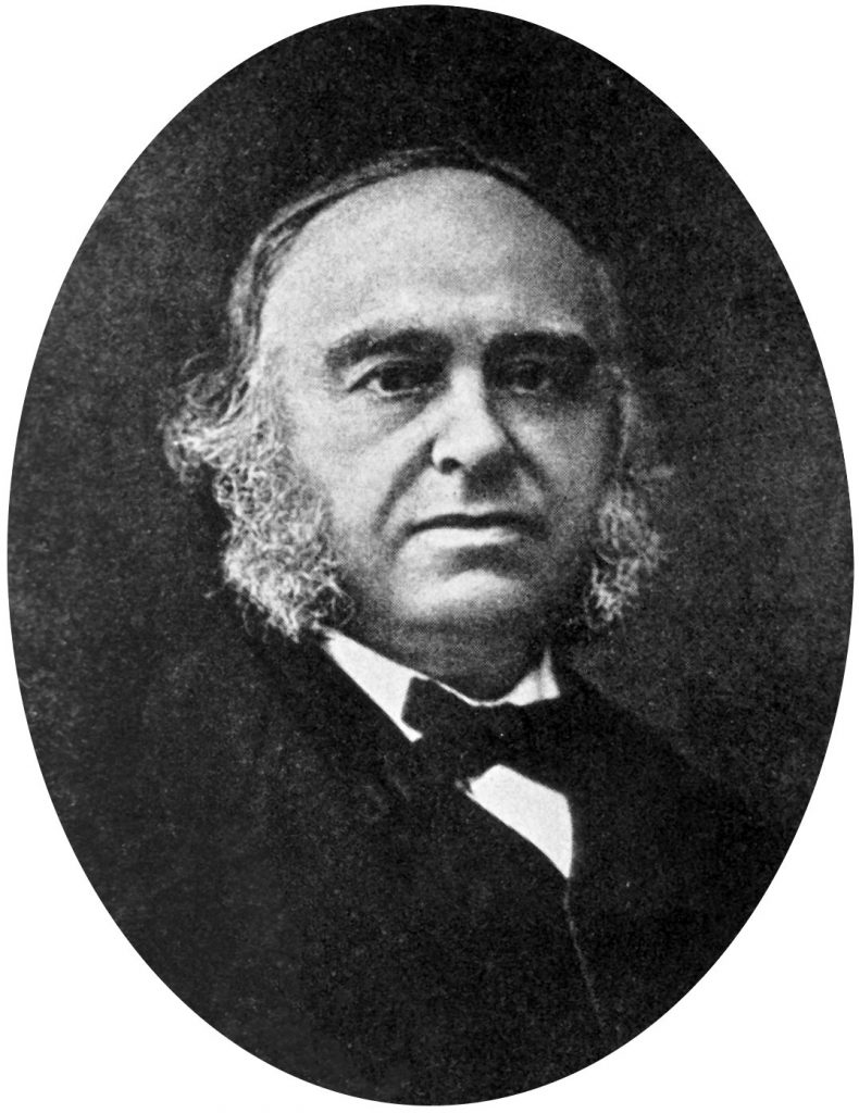
Localizationism
For hundreds of years, physicians attempted to correlate behaviors with changes in the brain. In the mid 1800s, the physician Paul Broca contributed to localization theory by concluding that specific areas of the brain were responsible for carrying out specific functions. This idea was supported by ablation studies that demonstrated that when different brain structures were ablated, or lesioned, there were specific associated functional losses. Further, electrically exciting specific brain structures resulted in eliciting specific behaviors.
Most likely, some behaviors are more localized than others, but still rely on signals from across many other brain areas. As with most fields of biology, absolutes are rare in neuroscience.
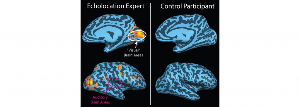
Plasticity
The real strength of our brain is its flexibility: brains are capable of changing and adapting to a wide variety of circumstances. Blind people use their visual areas of the brain while echolocating, stroke survivors can regain lost motor functions using the unaffected brain circuits, and babies can effortlessly learn two languages simultaneously in a bilingual household.
Plasticity is based on the idea that not only is the brain capable change, but that our experiences change the structure and function of our nervous system.
Key Takeaways
- Neuroscience is the study of the nervous system and is an integrative field of study that incorporates biology, psychology, chemistry, physics, mathematical modeling, and health care providers.
- The study of neuroscience is accomplished through experimental studies, case studies, and the use of experimental animal models.
- There are many popular myths concerning neuroscience and it is important to analyze data that refutes these myths.
- Though the field of neuroscience is relatively young and ever-changing, humans have been interested in the brain and its function for centuries.
Test Yourself!
Attributions
Portions of this chapter were remixed and revised from the following sources:
- Open Neuroscience Initiative by Austin Lim. The original work is licensed under a Creative Commons Attribution-NonCommercial 4.0 International License.
Media Attributions
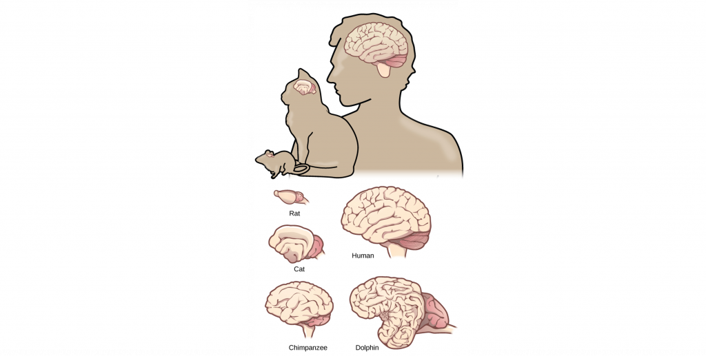
- Comparison of Brain Size Across Species © OpenStax adapted by Valerie Hedges is licensed under a CC BY-SA (Attribution ShareAlike) license
- Neuroscience is an integrative field of study © T. Wesley Mills adapted by Valerie Hedges is licensed under a Public Domain license
- Scientific method © Efbrazil adapted by Valerie Hedges is licensed under a CC BY-SA (Attribution ShareAlike) license
- Phineas Gage and his injury adapted by Valerie Hedges is licensed under a Public Domain license
- Commonly used animal models in neuroscience adapted by Valerie Hedges
- Preparations © Valerie Hedges is licensed under a CC BY-SA (Attribution ShareAlike) license
- Trepanated skull © Rama adapted by Valerie Hedges is licensed under a CC BY-SA (Attribution ShareAlike) license
- Paul Broca © Unknown is licensed under a Public Domain license
- Plasticity © Alan Thistle adapted by Valerie Hedges is licensed under a CC BY-SA (Attribution ShareAlike) license
The study of the nervous system
Electrically excitable cells of the nervous system that receive and transmit signals to different areas of the body.
Chemicals that are released by neurons or other cells that bind to receptors on other neurons or cells to elicit change in the target cell.
The electrical signal transmitted by the axon of a neuron.
Neurological disorder of motor function resulting from the loss of dopamine-producing cells in the substantia nigra
Electroencephalogram. A noninvasive method to record activity within the brain by using electrodes that are placed on the scalp.
Neurological disorder that causes seizures or unusual sensory experiences
a highly detailed description of a single patient and their condition
Performing an experiment in an intact, living organism, whether human or nonhuman
an experiment performed on cultured cells or isolated molecules of DNA, RNA, or protein
an experiment that takes a section of the living organism, such as a slice of brain, a tissue biopsy, or a detached frog leg
The process where new neurons are generated
A neural injury caused by either blockage of a blood vessel or bursting of a blood vessel within the brain
The ability to change
Neurodegenerative disease that causes cell death and dementia
specific areas of the brain were responsible for carrying out specific functions
A procedure that removes a structure and/or its function
