
59 Emotions: Fear
Nearly everyone has experienced the prototypical fear response: Imagine reading a book when you see a spider skittering along the wall. Suddenly, you’ll feel your heart racing, your breathing increase, dryness of your mouth, and your palms sweating. You probably won’t notice the dilation of your pupils or the change in liver activity and digestion. This sympathetic nervous system activity is downstream of being presented with a fearful stimulus. But upon closer inspection, it was no spider at all—just an errant piece of brown fuzz picked up by a draft. Within minutes, your body’s physiology returns back to normal.
This anecdote points out a few important features about the fear response. First, the onset of the fear response is quick, and so is the dissipation of the fear. Second, it is triggered by exposure to a perceived threat, regardless of whether the stimulus is a genuine threat or not (the overwhelming majority of spiders are clinically harmless to humans!). Third, the fear response is greatly modified by knowledge and experience—an entomologist would recognize that the spider is a harmless house spider and would, instead of fear, display curiosity, interest, boredom, or other emotions. On the other hand, someone who has been bitten by a dangerous spider and sent to the hospital when younger would have a much stronger physiological response.
Fear is likely the most evolutionarily ancient emotion, and is highly protective. When encountering a hungry mountain lion, faces displaying the traits of fear (enlarged eyes, flared nostrils, and a slightly open mouth accompanying a gasp) would signal to others nearby that a threat is nearby, which helps initiate heightened alertness and the appropriate fight-or-flight response.
Amygdala and Fear
Klüver-Bucy Syndrome
In 1939, two researchers named Heinrich Klüver and Paul Bucy described a unique set of emotional deficits in monkeys with bilateral temporal lobe removal, including removal of the, further providing evidence of the neuroanatomy of emotion. Most notably, the monkeys failed to display fear, including their facial expressions and vocalizations even in the face of life-threatening stimuli such as a large snake. Further, they also had decreased anger or aggression. They also display visual agnosia (the inability to recognize faces or objects visually), psychic blindness, hypersexuality, and hyperorality (an inappropriate fixation with using the mouth to interact with surroundings, such as licking or eating nonfoods). Collectively, these sets of symptoms are called Klüver-Bucy syndrome.
Although it is primarily an experimental manipulation observed in monkeys, some human patients may develop the condition, such in after brain surgery, stroke, viral encephalitis, traumatic brain injury, and many others. In addition to the symptoms described in Klüver-Bucy Syndrome, humans also display “flattened” emotions. The lesioning of the amygdala clearly results in decreased expression of fear. Stimulation of the amygdala, however, causes in increase in anxious, fearful, and aggressive responses in humans and other animals, such as piloerection in cats. In fact, amygdala activity is increased when shown images of fearful faces, but not neutral or happy faces when measured with fMRI.
Amygdala Lesions
Patient SM is a notable case study of a person who does not experience the fear response. Born with an extremely rare genetic condition called Urbach-Wiethe disease, Patient SM progressively developed calcification in her amygdala bilaterally, causing destruction and cell death. Like other people with Urbach-Wiethe disease, she had no severe significant cognitive deficits, except for the inability to experience fear.
In one test, she was shown a variety of emotionally charged videos, then asked to rate the intensity of each clip with respect to different emotions. SM found clips from America’s Funniest Home Videos to be just as funny as the control patients, and she found the clips of disgusting toilets to be just as repulsive. But, when presented with clips depicting ghost hauntings or suspenseful serial killers on the loose, SM rated these stimuli as being non fearful.
Other studies challenged Patient SM with more concrete threats. Researchers brought SM to a pet store, where she asked to handle the snakes. She was stopped by an employee before she put her hand into a tarantula cage out of curiosity. The researchers also took her to the Waverly Hills Sanatorium haunted house, where she bravely led a group of strangers through the house populated by actors dressed as monsters and ghosts. Although she did not display any fearful behaviors, such as hesitation to walk through the darkened corridors, patient SM reported the sensation of exhilaration and enthusiasm, akin to riding a roller coaster. She also was asked to recall some of her past real-life, fear-provoking experiences, such as when she was attacked in a domestic violence incident or was held up by a stranger at knife point in a public park. In none of these cases did she ever report feeling fear, although she was upset and angered at the situation. The destruction of her amygdala seemed to make her resilient against PTSD: the day after being threatened with a knife to her throat, she walked past the very same park bench.
Types of Fear
Scientists have long realized there are at least two distinct types of fear:
- Innate fear in which subjects avoid certain stimuli such as snakes or spiders even though they may never have seen them before
- Learned fear in which a stimulus or situation causes arousal or anxiety because it has been associated with a painful or negative experience in the past
There are two important protocols used to examine learned fear: fear conditioning and conditioned defeat.
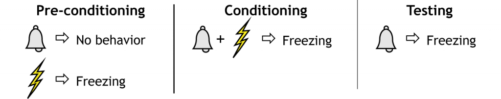
Fear Conditioning
One of the best studied laboratory models of fear comes from the work of Joseph LeDoux who studied the brain circuit that mediates learned or conditioned fear in laboratory rats. In these studies, LeDoux used a classical conditioning procedure to induce what he called “conditioned fear”. In classical conditioning (think Pavlov’s dogs), a neutral stimulus that normally would not cause any physiological response (called a conditioned stimulus, e.g., a ringing bell) is paired with a meaningful stimulus (called an unconditioned stimulus, e.g., the presence of food) that elicits a behavioral response (unconditioned response, e.g., drooling). Eventually, the behavior (drooling) occurs in response to the conditioned stimulus (bell) alone.
Instead of pairing the conditioned stimulus with a positive unconditioned stimulus like food, LeDoux paired the conditioned stimulus with an electrical shock. After pairing the shock to the stimulus multiple times, the animals responded to the conditioned stimulus alone (no shock) in the same way they did to the electrical shock alone. This is referred to as the conditioned emotional response.
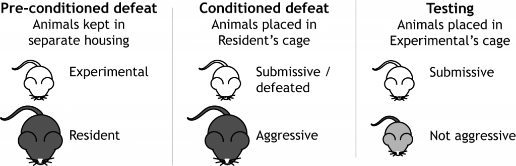
Conditioned Defeat
In this model the experimental subject (the animal whose behavior is being examined) is placed in the home cage of a larger, resident animal. This typically results in aggressive behaviors displayed by the resident animal toward the experimental subject. The resident animal will usually win the encounter because it is larger and in its own territory. The experimental animals will show submissive and defensive posturing and does not attack or threaten its opponent.
Experiencing this defeat has major long-lasting effects on the experimental subject. Following defeat, the animal rarely shows aggression even to non-aggressive animals placed in the subject’s home cage (an intruder would typically cause aggression). Because this paradigm has parallels with the earlier fear conditioning studies that paired an acoustical tone with electrical shock to the feet, it is often referred to as “conditioned defeat;” the experimental subject has been conditioned to respond to all other animals with submissive defensive postures.
Fear and Aggression
Fear and anger are two very closely related emotions. As with many other emotions, anger manifests in complex ways. The anger spectrum runs from low (irritation) to high (rage), and from quick (lashing out) to persistent (vengeful). Anger can manifest behaviorally as aggression, though clearly in humans we can feel angry without showing signs of aggression. In animal models, however, we can only judge anger through their display of aggressive behaviors.
Aggression is affected by circulating levels of androgens, as castration (removal of the testes) can reduce aggressive behavior. As with fear, amygdala activation is also related to aggressive displays, as bilateral removal of the amygdala causes animals to be less aggressive and more docile. A type of psychosurgery, amygdalectomy (bilateral surgical lesioning of the amygdala), can cause a “taming” effect in humans, and is still performed today, though rarely.
In addition to amygdala circuits, regions in the frontal cortex decrease activity during acts of aggression, suggesting that frontal circuits actively inhibit the limbic system, which drive our more “primitive” responses. Researchers in the 1920s performed a series of lesion experiments, systematically injuring different parts of the brain in cats. To their surprise, when the cortex of the cat was surgically separated from the rest of the nervous system, a procedure called the decorticate preparation, the cats would exhibit a hyper-aggressive response to stimuli. For example, an innocuous touch of the tail would trigger violent clawing, biting, and hissing, behaviors which were described as sham rage. The same sham rage behavior was observed following a decorticate preparation that also included the anterior hypothalamus. However, when the decorticate preparation included both the anterior and poster hypothalamus, sham rage was no longer observed. They concluded that rage (and other powerful emotions) is normally under inhibitory control by the cortex. Altered frontal cortical action may therefore account for one reason why two different people would react to the same anger-provoking stimulus in different ways.
In a separate set of experiments, cats had different areas of their hypothalamus electrically stimulated while being presented with a rat. These experiments indicated that stimulation of different areas of the hypothalamus elicits distinct displays of aggressive behavior. When the medial hypothalamus is electrically stimulated, the cat displays what is caused a “threat attack”. The threat attack includes threatening behaviors such as hissing, but the cat did not attack the rat. If the lateral hypothalamus was electrically stimulated, the cat displayed a “silent-biting attack”. In this case, the cats did not display exaggerated threatening behaviors, but rather the cat would quickly and viciously attack the rat’s neck.
Key Takeaways
- Lesions of the amygdala in monkeys leads to Klüver-Bucy Syndrome, which includes decreased expression of fear.
- Lesions of the amygdala in people results in decreased expression of fear, as demonstrated with patient SM.
- Learned fear can be studied using either fear conditioning or conditioned defeat tests.
- Fear conditioning pairs a neutral stimulus, like a light or a tone, to a harmful stimulus, like a shock.
- Conditioned defeat submits an experimental animal to aggressive behaviors from another animal, after which the experimental animal will continuously show submissive behaviors to others.
Attributions
Portions of this chapter were remixed and revised from the following sources:
- Foundations of Neuroscience by Casey Henley. The original work is licensed under a Creative Commons Attribution-NonCommercial-ShareAlike 4.0 International License
- Open Neuroscience Initiative by Austin Lim. The original work is licensed under a Creative Commons Attribution-NonCommercial 4.0 International License.
Media Attributions
- Fear Conditioning © Casey Henley is licensed under a CC BY-NC-SA (Attribution NonCommercial ShareAlike) license
- Conditioned Defeat © Casey Henley
'Fight or Flight' system
