
58 Emotions: Overview
Emotions
At the airport, you can observe people experiencing a wide range of emotions: Sadness at seeing family members off, fear and anxiety for those about to fly for the first time, love when a long-distance relationship is reunited, and anger over unpredictable cancellations. Emotions are complex neurophysiological states that contribute to an internal feeling and guide behavior. Some emotions are pleasant (joy), some are negative (disgust), and some are a mix of both (nostalgia). Some are short-lasting (surprise) while others may persist over years (vengefulness). However, beyond this statement, it becomes very difficult to put a clear-cut definition on “emotion”. The difficulty with defining emotion arises because of the fluid nature of emotions: they exist on a spectrum, multiple emotions are experienced simultaneously, each emotion is perceived by different people in unique ways, and everyone has a slightly different interpretation and understanding of an emotion.
The field of affective neuroscience seeks to understand the neural mechanisms that underlie emotion. The field has expanded with the help of functional imaging methods like EEG and fMRI, where changes in brain activity can be measured and quantified as a person experiences different emotion-provoking stimuli. Affective neuroscientists work to develop biology supported therapies for disorders such as depression, PTSD, and addiction, which are dysregulations of the emotions of sadness, fear, and desire, respectively.
Faces of Emotion
Although best known for his theory on evolution, naturalist Charles Darwin published prolifically about other topics in biology, ranging from botany, coral reefs, and even a treatise on psychology. In this 1872 text, called The Expression of Emotions in Man and Animals, Darwin suggested that similar emotional responding is found across different cultures, and to some extent, even in nonhumans. In his view, the main purpose of emotive expression is to communicate survival cues between individuals: a relaxed expression conveys safety, while a fearful expression promotes alertness, since danger may be nearby. Darwin also suggested that we gain survival information from non-human behaviors, for example, a hissing snake or a snarling lion is an immediate threat that should cause fear or other avoidance behaviors.
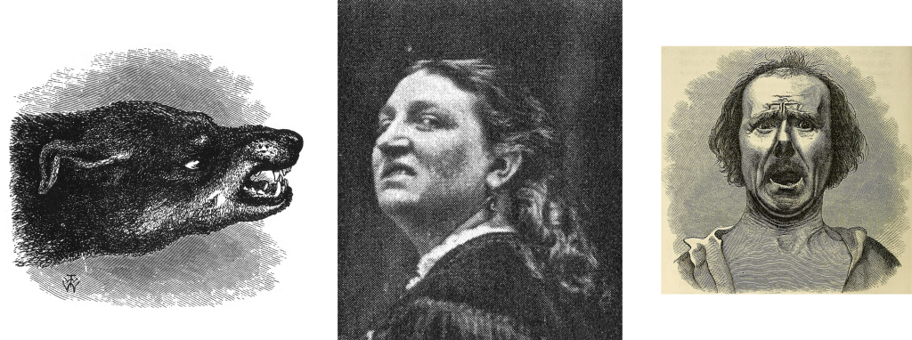
Moving forward into more modern times, the American psychologist Paul Ekman expanded on Darwin’s theory, distilling down the range of human emotions to belonging in one of seven basic categories of emotions: anger, contempt, disgust, fear, happiness, sadness, and surprise. If the purpose of emotion was to communicate pro-survival cues, then Ekman theorized that all humans, regardless of culture, would use similar facial expressions.
To test this hypothesis, Ekman visited a remote village in Papua New Guinea, where he studied a population that is isolated from any other known cultures. As predicted, these people made the same facial responses in reaction to various emotional circumstances. In 1972, Ekman published his theory of universal facial expressions.
Part of Ekman’s research led their team to develop a series of photographs of actors portraying six major emotions. This Ekman 60 faces (EK-60F) test has since been used to assess facial emotions with fascinating results: People with major depressive disorder or borderline personality disorder have a lessened ability to detect happiness in others, seeing emotional faces results in a similar emotion in the viewer, and people with dementia or Parkinson’s disease identify emotions as being less intense. Ekman’s research led them to develop the Facial Action Coding System (FACS), a system that uses facial anatomy to differentiate the features that are characteristic of different expressions. For example, a feature of a happy face is the flexing of the zygomaticus major and orbicularis oculi muscles, which produces an upward turn of the corners of the mouth and a rising of the cheeks. Other facial features, such as head movement, eye movement, and larger physical movements are also scored, and are also used to help identify emotions. The FACS can be used to formally describe why some smiles appear as genuine (a Duchenne smile, with simultaneous muscle action) while others look fake or forced (a non-Duchenne smile, characterized by the turn of the corners of the mouth without much change to the top part of the face).
History of Emotion Research
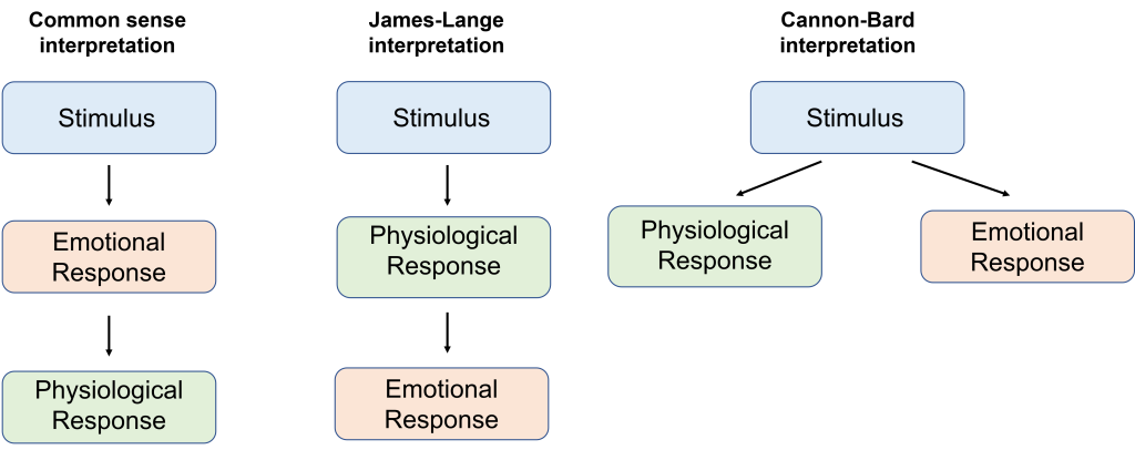
One of the older theories about the origin of emotion is based on the most common sense interpretation of cause and effect. For example, imagine some noticeable emotional stimulus, such as encountering a hungry lion on the sidewalk. The logical cause and effect explanation suggests that seeing the lion prompts the emotion of fear, which then causes the sympathetic nervous system “fight-or-flight” response (elevated heart rate and blood pressure, increased respiration, and cellular mobilization of energy). In the 1880s, psychologist William James and physician Carl Lange independently developed a new theory about the origin of emotion. According to the James-Lange theory of emotion, and contrary to a common sense understanding of the origin of emotion, the body’s physiological changes precede the onset of an emotional response. For example, imagine encountering that same hungry lion on the sidewalk. The James-Lange theory tells us that the perception of the threat of being eaten causes the sympathetic nervous system response, and that these physiological changes trigger the onset of fear.
Soon after, in the 1920s and 1930s, two physiologists named Walter Cannon and his doctoral student Philip Bard criticized the James Lange theory. In one experiment, they surgically removed the entire sympathetic nervous system from cats, destroying the nerves that regulate vascular dilation, the activity of liver enzymes, and the reaction that causes the hair standing on end. These cats were then put before a threatening aggressor. If the James-Lange theory was true, then the physiological changes should precede the emotive response. However, the cats exhibited the fear / aggression response (such as posturing, hissing, and clawing) even without an intact sympathetic nervous system. Relatedly, patients with spinal cord injuries have a similar lack of autonomic inputs to the brain, but their capacity for emotional responding is still intact.
In a second criticism, Cannon and Bard proposed that the physiological changes seen in sympathetic nervous system activity may arise for a variety of reasons, not always for emotionally salient reasons. For example, intense exercise causes strong cardiorespiratory changes; however, we do not necessarily feel a strong emotional state after this physiological perturbation. Likewise, exogenous administration of epinephrine, onset of fever, or being in cold temperatures may also trigger some physiological changes without causing a strong emotional response. Based on their evidence opposing the James-Lange theory, Cannon and Bard developed an alternative explanation for the origin of emotions. According to the Cannon-Bard theory of emotion, the perception of an emotionally charged stimulus prompts simultaneous but independent activation of both the autonomic nervous system and the emotional response. The Cannon-Bard theory also draws attention to the neuroanatomical structures that trigger the autonomic and emotional responses, with a particular focus on diencephalon structures such as hypothalamus and thalamus.
Papez Circuit
Just years later, in 1937, American neuroanatomist James Papez (pronounced payps) ascribed emotional behavior to a particular series of brain structures. These structures, collectively called the Papez circuit, consist of the hypothalamus, cingulate gyrus, thalamus, hippocampus, and more. The model that Papez developed started with a stimulus that activates the thalamus, which in turn activates a loop of activity between the hippocampus, hypothalamus, thalamus and cortex to produce both a physiological response and emotional feelings.
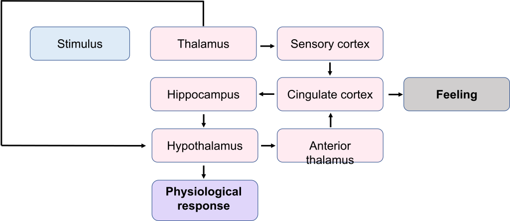
Papez observed unusual aggression among animals with injury to these structures, suggesting that emotional responding is distributed across many areas, rather than localized. (Notably missing from the Papez circuit structures is the amygdala, which was later added in a future revision in the 1950’s.)
Structures Involved in Emotion
Emotions are one of the most holistic functions of the brain, incorporating neural circuitry from across several different structures, ranging from the phylogenetically older to the most advanced frontal cortical areas. The structures of the Papez circuit are not a comprehensive list of brain structures involved in emotional processing but offer a good starting place for describing the anatomy of emotion.
Amygdala
A limbic structure called the amygdala contributes heavily to processing the valence of emotional experiences. Roughly the shape and size of an almond, the amygdala is part of the temporal lobe. Generally, the amygdala is subdivided into several nuclei, including the basolateral amygdala, central nucleus, and cortical nucleus. As a temporal lobe structure, the amygdala is strongly implicated in emotional memory formation. Emotional memories can be either positive valence (such as the happiness you may have experienced at a birthday party when younger) or negative valence (such as childhood trauma or being teased as a child).
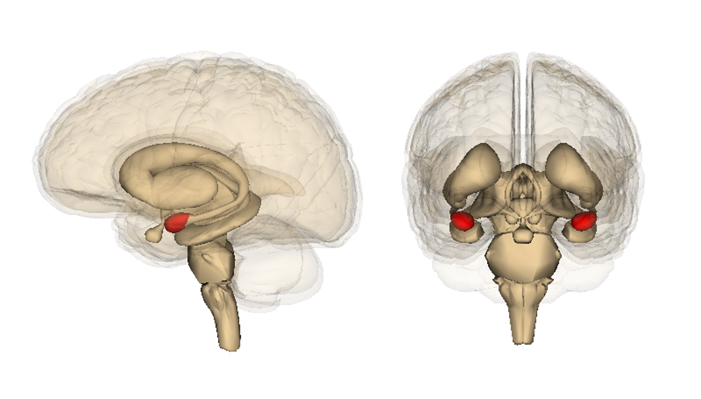
Of the forms of declarative memory, autobiographical memories more often have emotional content compared to semantic memories. Amygdala lesions have been used as a last resort treatment for patients with temporal lobe epilepsy or psychiatric conditions with pathological and dangerous aggressiveness. These psychosurgery strategies have been variably successful, but they often have high complication rates and upwards of a 4% mortality rate. These treatments are rarely used today.
Hypothalamus
One of the major output signaling pathways of the amygdala is the hypothalamus, found at the base of the brain. The hypothalamus is often seen as the neural structure that initiates endocrine responses in the rest of the body, such as hormone production. Several different behaviors, ranging from homeostasis, hunger, and circadian regulation are modulated by hypothalamic signals.
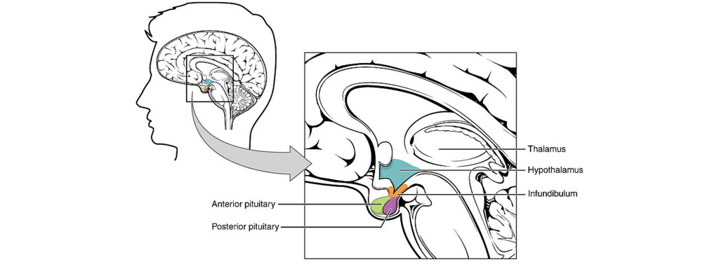
Pituitary Gland
Downstream from the hypothalamus is the pituitary gland, a pea-sized endocrine organ that protrudes from the base of the brain. It is strongly involved in the production and release of neurohormones, signaling molecules produced by nerve cells that travel throughout the bloodstream to influence the activity of several organs throughout the entire body. Recall that the pituitary gland is subdivided into two regions with distinct anatomical and functional differences. Hypothalamic magnocelluar neurons produce the neurohormones oxytocin and vasopressin. The axons of these neurons project to the posterior pituitary gland, which then releases these hormones into the bloodstream.
Oxytocin plays a significant role in the development and maintenance of prosocial behaviors, acts such as trust, compassion, and empathy—all actions that enhance interpersonal relationships. Interestingly, while oxytocin generally strengthens the social bonds between people, it promotes antisocial behaviors against those not perceived to be within one’s own social group. Disorders of the oxytocin system are believed to contribute to autism spectrum disorder and psychopathy, two complex conditions characterized partly by social impairment. Some studies have examined the therapeutic use of nasal oxytocin for a variety of psychiatric conditions, but the studies have been unable to demonstrate strong clinical effects despite success in nonhuman animal models.
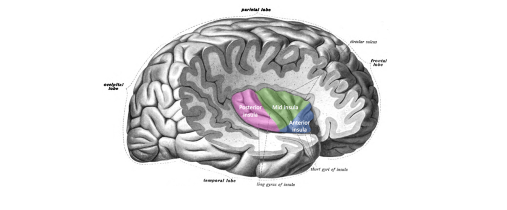
Insula
The insula (or insular cortex) is the lobe of the cortex buried deep within the lateral fissure. Although not visible from a side view, the insula is often considered to be the fifth lobe of the telencephalon. Early studies by Wilder Penfield where he directly stimulated the brain during open brain surgery led researchers to suggest that the insula contributes to interoception, detecting the internal state of the body and conveying that information for processing.
In functional imaging studies, the insula is involved in the recall or many different emotional stimuli, especially those emotions that have a sensory component. Notably, the insula is strongly implicated in the emotion disgust. For example, a patient is placed in an fMRI scanner while breathing through a mask, which allows the experimenters to change the smells that are perceived. Patients are then given pleasant smells (such as passion fruit, pear, or mint), a neutral smell, or unpleasant smells (like ethyl-mercaptan or isovaleric acid, which smells like skunk or body odor, respectively). There is increased activity of the anterior insula in response to the unpleasant smells but not the pleasant smells.
The insula also responds to social cues related to disgust as well. When a patient in an fMRI sees a video of a person smelling something unpleasant and reacting with a “repulsed” face (the closing of the nostrils and curling of the upper lip), their anterior insula likewise increases in activity just as if they had smelled it themselves. In addition to sensory stimuli, feelings of social repugnance (unwarranted violence, murder) or moral disgust (incest) also increase insula activity. Atypical insula activity is implicated in behavioral disorders. For example, insensitivity to disgust can lead to squalor-dwelling conditions (sometimes seen in excessive hoarding or late cognitive decline), which puts those people at heightened health risks due to regular exposure to unsanitary conditions. Substance use disorders, PTSD, and suicide attempts have all been associated with atypical insula activity.
Key Takeaways
- Affective neuroscience studies emotions.
- There are seven basic classes of emotions: Anger, contempt, disgust, fear, happiness, sadness, and surprise.
- The Papez circuit relates several brain structures to emotional feeling and physiological feelings, including the amygdala, hypothalamus, pituitary gland, and insula.
Attributions
Portions of this chapter were remixed and revised from the following sources:
- Open Neuroscience Initiative by Austin Lim. The original work is licensed under a Creative Commons Attribution-NonCommercial 4.0 International License.
Media Attributions
- Darwin Emotions © Charles Darwin adapted by Valerie Hedges is licensed under a Public Domain license
- Emotion interpretations © Valerie Hedges is licensed under a CC BY-NC-SA (Attribution NonCommercial ShareAlike) license
- Papez circuit © Valerie Hedges is licensed under a CC BY-NC-SA (Attribution NonCommercial ShareAlike) license
- Amygdala © Life Science Databases adapted by Valerie Hedges is licensed under a CC BY-SA (Attribution ShareAlike) license
- Hypothalamus © OpenStax College adapted by Valerie Hedges is licensed under a CC BY (Attribution) license
- Insula © Schappelle adapted by Valerie Hedges is licensed under a CC BY-SA (Attribution ShareAlike) license
'Fight or Flight' system
memory that involves remembering events and facts. Also called explicit memory.

.png https://commons.wikimedia.org/wiki/File:The_expression_of_the_emotions_in_man_and_animals_(1872)_(14771346374).jpg)