
53 Sex Steroid Hormones
Control of gonadal hormone release relies on activation of the hypothalamic-pituitary-gonadal (HPG) axis. Gonadal hormones are important for development of the body and brain, changes during puberty, and the activation of some behavior in adulthood like reproductive behavior and aggression.
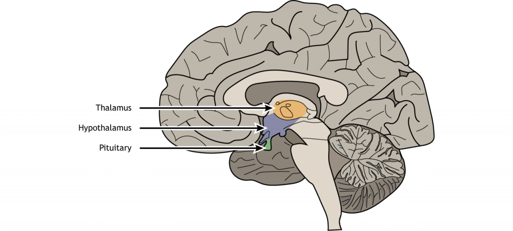
Hypothalamus
As a refresher, the hypothalamus, which is located inferior to the thalamus, integrates information from many regions of the central nervous system and maintains homeostasis in the body. The hypothalamic regulation of gonadal hormones and sex behavior is managed via hormone release by the pituitary gland.
View the hypothalamus using the BrainFacts.org 3D Brain
View the pituitary using the BrainFacts.org 3D Brain
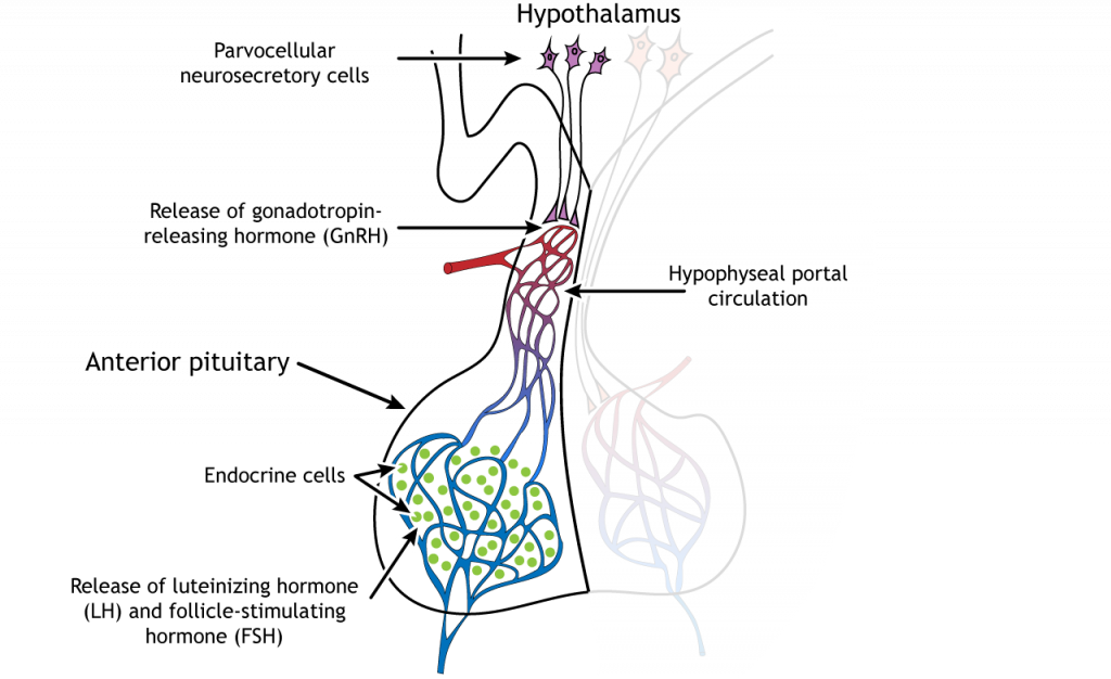
Hormone Release
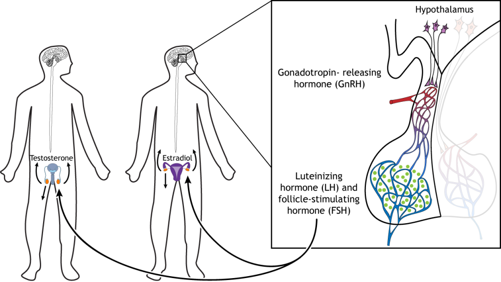
Gonadal hormone release relies on anterior pituitary function. In the hypothalamus, the parvocellular neurosecretory cells release a hormone called gonadotropin-releasing hormone (GnRH) into the hypophyseal portal circulation. When GnRH reaches the anterior pituitary, it causes the endocrine cells of the pituitary to release luteinizing hormone (LH) and follicle-stimulating hormone (FSH) into the general circulation.
The LH and FSH travel through the circulatory system and can act on the gonads, either the testes in males or ovaries in females. In response to the pituitary hormones, the testes release testosterone, an androgen, and the ovaries release estradiol, an estrogen, into the blood stream. After puberty, the LH and FSH are also critical for the maturation of sperm and egg cells.
Sex Steroid Hormone Synthesis
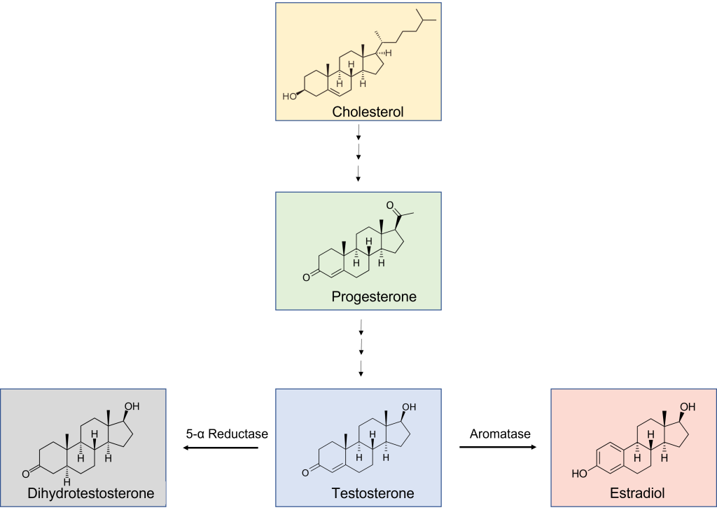
Sex steroid hormones are produced by the gonads (ovaries or testes). All sex steroid hormones are originally generated from cholesterol. Males typically have higher concentrations of androgen sex steroid hormones, such as testosterone and 5-alpha-dihydrotestosterone (DHT). Androgens are primarily released from the testes. Females typically have higher concentrations of estrogens (such as estradiol) and progesterone that are primarily released from the ovaries. Importantly, males also have estrogens and progesterone, and females have androgens. The only difference is the relative concentration between males and females.
In the synthesis pathway, enzymes are responsible for converting one hormone into another hormone. The enzyme 5-α reductase converts testosterone into DHT. DHT is important for masculinization of the body structures. Through just one chemical reaction, the enzyme aromatase converts testosterone into estradiol.
Hormone Action
Once the gonadal hormones are synthesized and enter the circulation, they are able to act on cells that express either androgen receptors or estrogen receptors. Testosterone and estradiol are steroid hormones that can affect target cells through two different mechanisms:
- Classical mechanism: Steroid hormones like testosterone and estradiol are able to pass through the phospholipid membrane of a neuron because they are lipophilic. Some neurons express receptors for these hormones. Androgen receptors bind androgens like testosterone while estrogen receptors bind estrogens like estradiol. When a hormone binds to a receptor in the neuron, the hormone-receptor complex dimerizes and moves into the nucleus where it can bind to specific promoter sites on the DNA and act as a transcription factor to turn on or off certain genes (genomic effects).
- Rapid mechanism: Sex steroid hormones can also bind to membrane-bound receptors and can exert more rapid effects within the cell.
Animation 52.1. Steroid hormones can cross the phospholipid bilayer and bind to hormone receptors. The receptors dimerize, move to the nucleus, and interact with DNA, altering transcription of certain genes. ‘Estradiol Action’ by Casey Henley is licensed under a Creative Commons Attribution Non-Commercial Share-Alike (CC BY-NC-SA) 4.0 International License.
The testes secrete testosterone during the prenatal critical period. The effect of this testosterone is to masculinize and defeminize the brain, body, and behavior, and this is accomplished through the transcription of a specific set of genes. However, many of those genes are not transcribed by the action of androgen receptors interacting with the DNA. Instead, when testosterone enters the cell, it does not always bind to androgen receptors. Some neurons also express the enzymes 5-α reductase and aromatase that break testosterone down into its metabolites: DHT and estradiol.
Animation 52.2. After testosterone enters the cell, if it does not bind to an androgen receptor, it can be metabolized by enzymes in the cell. 5-α reductase converts testosterone into dihydrotestosterone. DHT, like testosterone, can bind to and activate androgen receptors. The enzyme aromatase converts testosterone into estradiol. Estradiol is an estrogen and can bind to and activate estrogen receptors. During developmental critical periods, the aromatization of testosterone to estradiol leads to the transcription of masculinizing genes in some animals like rodents. ‘Aromatization’ by Casey Henley is licensed under a Creative Commons Attribution Non-Commercial Share-Alike (CC BY-NC-SA) 4.0 International License.
In some mammals, like rodents, this conversion of testosterone to estradiol through the actions of the enzyme aromatase is the main process by which neurons and the brain are masculinized. The estrogen receptors cause the transcription of masculinizing genes. Therefore, somewhat surprisingly, even though estrogen is typically thought of as a ‘female’ hormone, its actions during development are responsible for much of the masculinization that occurs in the brain in some animals.
The brain is by default “female” during development, and requires the testosterone secreted by the testes and converted into estradiol for “male” development. It should be noted, though, that estrogen does not appear to have these same masculinizing effects during human development.
Key Takeaways
- The hypothalamus directly controls release of gonadal hormones by controlling hormone release from the anterior pituitary.
- The hypothalamus releases gonadotropin-releasing hormone (GnRH).
- The anterior pituitary releases luteinizing hormone (LH) and follicle-stimulating hormone (FSH).
- The ovaries release estradiol.
- The testes release testosterone.
- Gonadal hormones bind to receptors and alter DNA transcription.
Attributions
Portions of this chapter were remixed and revised from the following sources:
- Foundations of Neuroscience by Casey Henley. The original work is licensed under a Creative Commons Attribution-NonCommercial-ShareAlike 4.0 International License
Media Attributions
- Hypothalamus Pituitary © Casey Henley is licensed under a CC BY-NC-SA (Attribution NonCommercial ShareAlike) license
- LH and FSH Release © Casey Henley is licensed under a CC BY-NC-SA (Attribution NonCommercial ShareAlike) license
- HPG Axis © Casey Henley is licensed under a CC BY-NC-SA (Attribution NonCommercial ShareAlike) license
- Sex steroid hormone synthesis © BorisTM, Rhododendronbusch, Klaus Hoffmeier, NEUROtiker, Aethyta adapted by Valerie Hedges is licensed under a Public Domain license
Below; toward the feet
enzyme that converts testosterone into DHT
enzyme that converts testosterone into estradiol
'lipid loving', combines with or dissolves with lipids/fats
