
1 The Neuron
Casey Henley
Neurons are the basic units of the brain. Their main function is to send electrical signals over short and long distances in the body, and they are electrically and chemically excitable. The function of the neuron is dependent on the structure of the neuron. The typical neuron consists of the dendrites, cell body, axon (including the axon hillock), and presynaptic terminal.
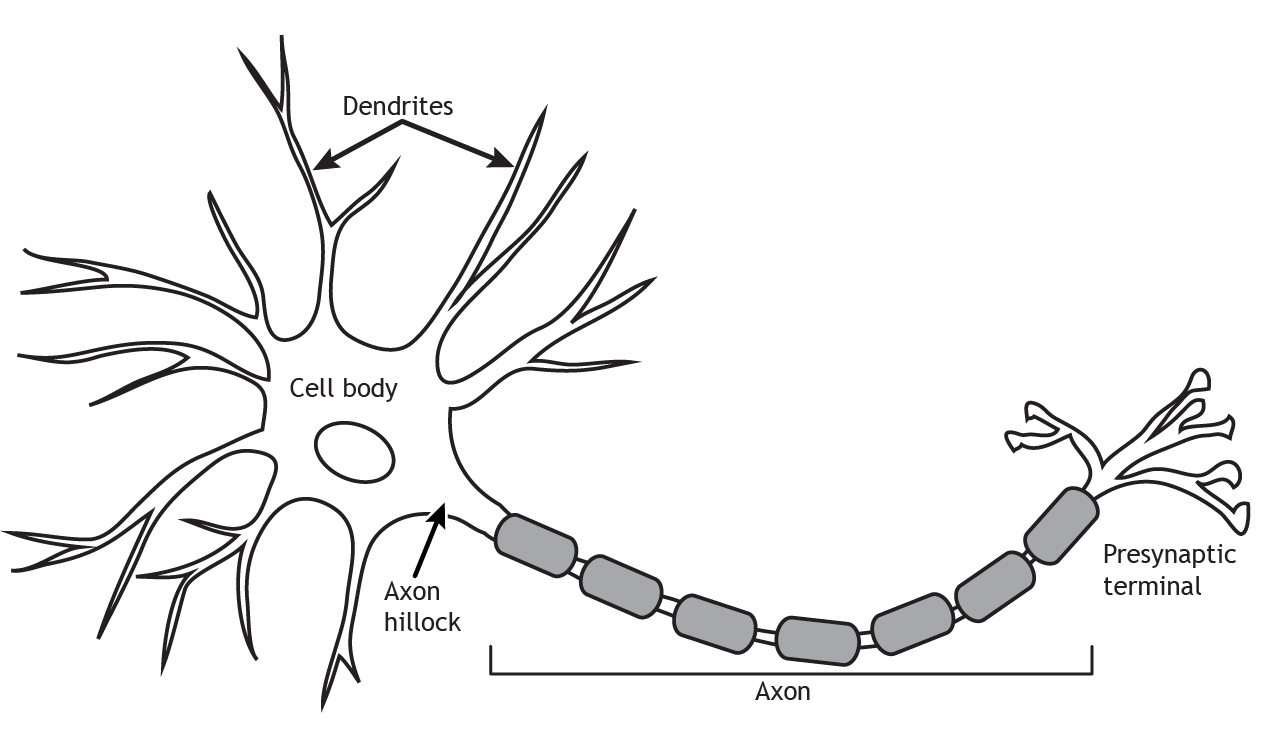
The structure of the neuron affects how it functions
Dendrites
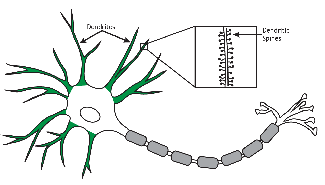
Dendrites, shown here in green, are processes that branch out in a tree-like fashion from the cell body. They are the main target for incoming signals received from other cells. The number of inputs a neuron receives depends on the complexity of the dendritic branching. Dendrites may also have small protrusions along the branches known as spines. Spines, illustrated in the inset box, are the sites of some synaptic contacts. Spines increase the surface area of the dendritic arbor, which may be an important factor in receiving communication.
Cell Body
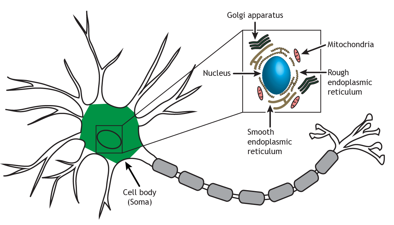
The cell body, shown here in green and also known as the soma, contains the nucleus and cellular organelles, including endoplasmic reticulum, Golgi apparatus, mitochondria, ribosomes, and secretory vesicles. The nucleus houses the DNA of the cell, which is the template for all proteins synthesized in the cell. The organelles, illustrated in the inset box, in the soma are responsible for cellular mechanisms like protein synthesis, packaging of molecules, and cellular respiration.
Axon
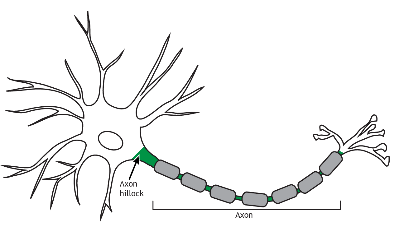
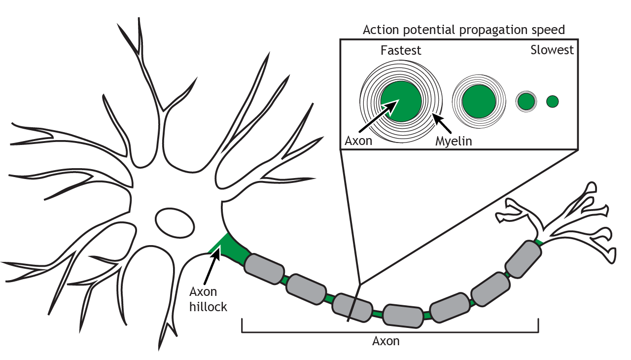
The axon, highlighted in green, is usually a long, single process that begins at the axon hillock and extends out from the cell body. The axon hillock is located where the cell body transitions into the axon. Axons can branch in order to communicate with more than one target cell.
Action Potential
The axon transmits an electrical signal, called an action potential, from the axon hillock to the presynaptic terminal where the electrical signal will result in a release of chemical neurotransmitters to communicate with the next cell. The action potential is a very brief change in the electrical potential, which is the difference in charge between the inside and outside of the cell. During the action potential, the electrical potential across the membrane moves from a negative value to a positive value and back.
Myelin
Many axons are also covered by a myelin sheath, a fatty substance that wraps around portions of the axon and increases action potential speed. There are breaks between the myelin segments called Nodes of Ranvier, and this uncovered region of the membrane regenerates the action potential as it propagates down the axon in a process called saltatory conduction. There is a high concentration of voltage-gated ion channels, which are necessary for the action potential to occur, in the Nodes of Ranvier.

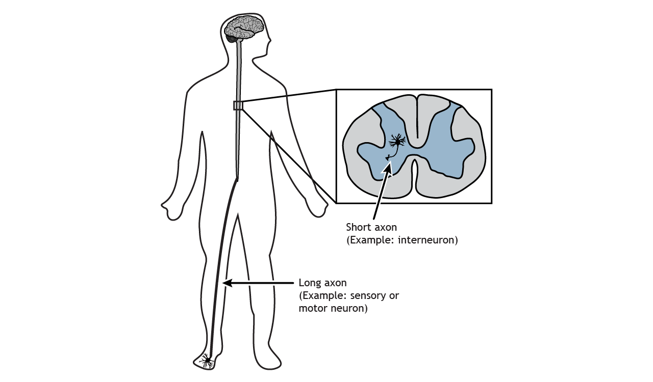
Axon Length
The length of an axon is variable depending on the location of the neuron and its function. The axon of a sensory neuron in your big toe needs to travel from your foot up to your spinal cord, whereas an interneuron in your spinal cord may only be a few hundred micrometers in length.
Axon Diameter
Axon diameter is also variable and can be used to differentiate different types of neurons. The diameter affects the speed at which the action potential will propagate. The larger the diameter, the faster the signal can travel. Additionally, larger diameter axons tend to have thicker myelin.
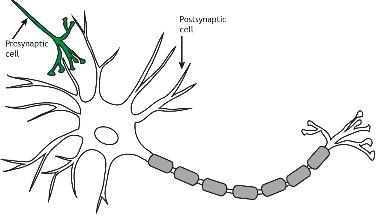
Presynaptic Terminal
The axon terminates at the presynaptic terminal or terminal bouton. The terminal of the presynaptic cell forms a synapse with another neuron or cell, known as the postsynaptic cell. When the action potential reaches the presynaptic terminal, the neuron releases neurotransmitters into the synapse. The neurotransmitters act on the postsynaptic cell. Therefore, neuronal communication requires both an electrical signal (the action potential) and a chemical signal (the neurotransmitter). Most commonly, presynaptic terminals contact dendrites, but terminals can also communicate with cell bodies or even axons. Neurons can also synapse on non-neuronal cells such as muscle cells or glands.
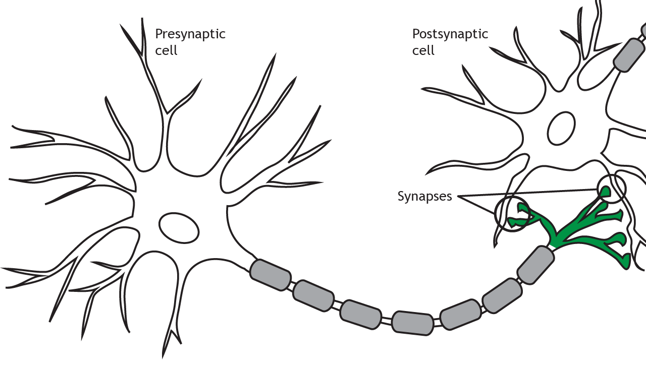
The terms presynaptic and postsynaptic are in reference to which neuron is releasing neurotransmitters and which is receiving them. Presynaptic cells release neurotransmitters into the synapse and those neurotransmitters act on the postsynaptic cell.
Variations in Structure
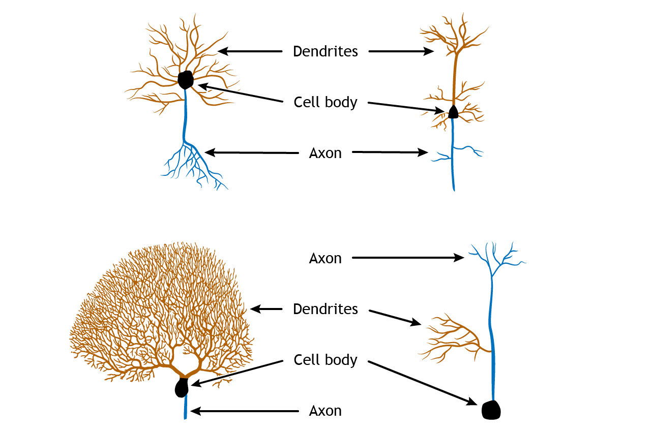
Although these typical structural components can be seen in all neurons, the overall structure can vary drastically depending on the location and function of the neuron. Some neurons, called unipolar, have only one branch from the cell body, and the dendrites and axon terminals project from it. Others, called bipolar, have one axonal branch and one dendritic branch. Multipolar neurons can have many processes branching from the cell body. Additionally, each of the projections can take many forms, with different branching characteristics. The common features of cell body, dendrites, and axon, though, are common among all neurons.
Conclusion
The neuron’s structure is essential to its function, from receiving inputs to transmitting signals. Understanding these basics lays the groundwork for exploring the complex networks that drive behavior and cognition.
Key Takeaways
- Neurons are the fundamental units of the brain, designed to transmit information via electrical and chemical signals
- The main structures of a neuron—dendrites, soma, axon, and presynaptic terminal—each play a critical role in communication
- Overall structure of the cell can vary depending on location and function of the neuron
Test Yourself!
Try the quiz more than once to get different questions!
- From memory, draw a neuron and identify the following structures: dendrites, soma, axon hillock, axon, myelin, nodes of Ranvier, presynaptic terminal.
- Describe functions of each neuronal structure depicted in your illustration.
- Predict what would happen to neuron function if myelin was destroyed
Video Lecture
