
44 Execution of Movement
Motor Cortex
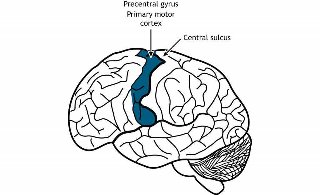
Once the plan for movement has been created, the primary motor cortex is responsible for the execution of that action. The primary motor cortex lies just anterior to the primary somatosensory cortex in the precentral gyrus located in the frontal lobe.
Primary motor cortex (M1) is a major motor control center, required for deliberate, voluntary movements, movements made in response to a “command.” Furthermore, motor cortex cells influence motor neurons, neurons located in the spinal cord that ultimately communicate with muscles or glands. This connection is so strong that the motor cortex cells are also called upper motor neurons. In this terminology, a lower motor neuron is found at the brain stem or spinal cord, and fires whenever the upper motor neuron sends a signal.
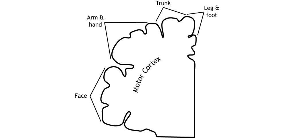
Like the somatosensory cortex, the motor cortex is organized by a somatotopic map. In the 1930s, neurosurgeon Wilder Penfield conducted several brain operations to treat patients with severe epilepsy. However, since the brain has no pain receptors, he was able to remove a portion of the skull under local anesthesia while the patients were awake and responding to his questions. During surgery, the goal was to electrically stimulate portions of the cortex to determine the origin of the seizures, and to ensure that areas critical to speech and hand movement were left untouched so that the patient would not have major impairments following surgery. This is currently done in neuro-oncology to reduce the loss of critical motor function and overall morbidity. Penfield progressively moved across different brain areas of M1 while using an electrode to stimulate patches of the cortex.
He had two major observations. First, stimulation caused contralateral activity: that is, stimulating the left side of the brain affects the muscle activity of the right side of the body. Secondly, by systematically moving across M1, he observed that different populations of neurons are responsible for communicating with specific muscle groups. For instance, dorsal M1 activates hip and trunk muscles, while more lateral M1 activates muscles of the face. Penfield discovered that within the motor cortex, different muscle groups are laid out in a rough topography, meaning that neurons that control the thumb are near other neurons that control the thumb, and near other populations of neurons that control the index finger. A graphical representation of this map is called the “motor homunculus,” in which body parts with larger representations in the brain are shown with larger size (much like the sensory homunculus).
Interestingly, the motor cortex does not map onto the body in such an exact way as does the somatosensory system. It is believed that upper motor neurons in the motor cortex control multiple lower motor neurons in the spinal cord that innervate multiple muscles. This results in activation of an upper motor neuron causing excitation or inhibition in different neurons at once, indicating that the primary motor cortex is responsible for movements and not simply activation of one muscle. Stimulation of motor neurons in monkeys can lead to complex motions like bringing the hand to the mouth or moving into a defensive position (Graziano et al, 2005).
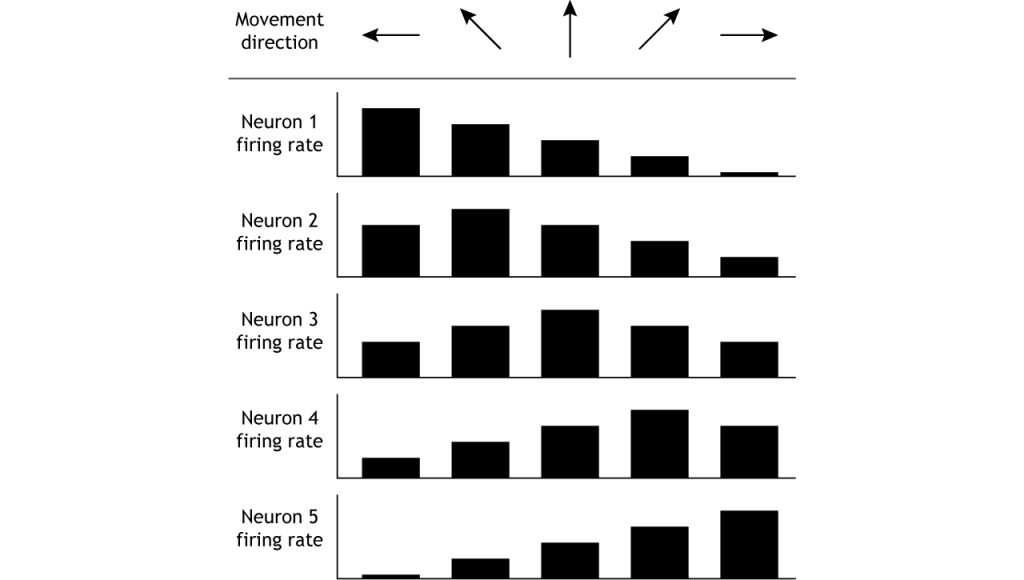
Population Coding
The motor cortex controls movement by using population coding mechanisms. Upper motor neurons are broadly tuned to a certain movement in a certain direction, meaning firing rate is highest when moving in one direction, but firing also occurs when moving in nearby directions. For example, when a monkey is trained to move its hand toward the left, neurons “tuned” toward left movement will be active immediately before and during the movement. Neurons tuned to other directions will also be active but at lower rates (Georgopoulos, et al, 1982). This means that the firing rate of one specific neuron does not give enough information to know direction of movement. It is the combined firing rates of an entire population of neurons that indicates direction.
Descending Spinal Tracts
There are multiple descending tracts within the spinal cord that send information from the brain to the motor neurons in the ventral horn. The lateral tracts are responsible for carrying information about voluntary movement of the arms and legs. The ventromedial pathways are responsible for carrying information about posture and balance.
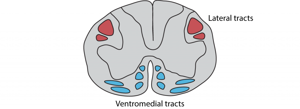
Lateral Tracts
The lateral pathways allow for voluntary movement of muscles and are under direct cortical control.
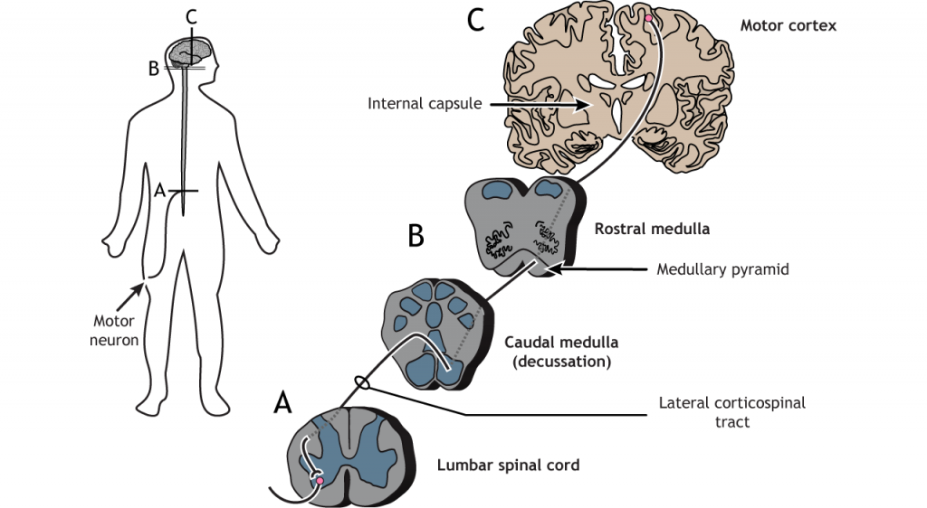
Corticospinal Tract
The largest of the lateral pathways is the corticospinal tract. This pathway sends information directly from the motor and premotor cortices down to the motor neurons in the spinal cord. Cortical axons travel through the brainstem and then cross the midline at the base of the medulla; like the somatosensory system, the right side of the cortex processes information for the left side of the body and vice versa. In the spinal cord, the axons travel through the lateral column and synapse in the ventral horn on motor neurons that typically innervate distal muscles.
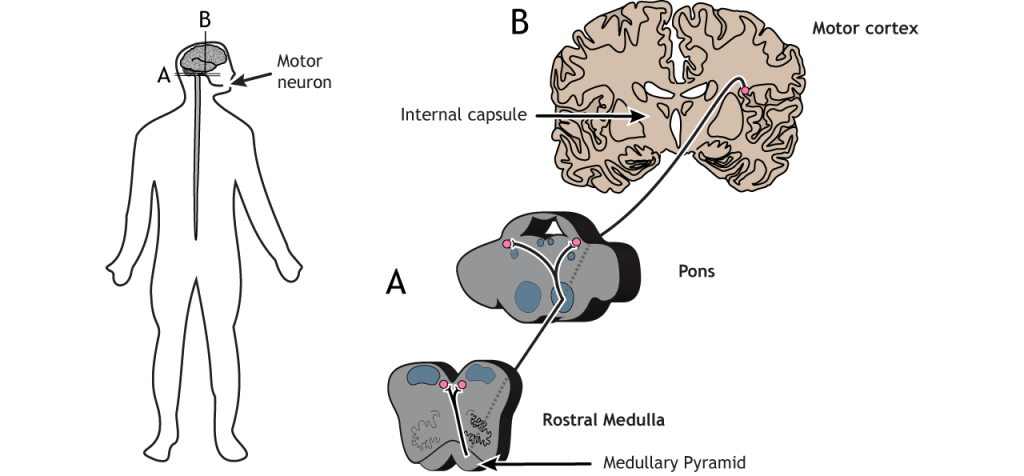
Corticobulbar Tract
The corticobulbar tract is another lateral tract and sends motor information to cranial nerves for motor control of the face. This path travels ipsilateral from the cortex into the brainstem where it branches off at the appropriate cranial nerve level in either the pons or the medulla and then innervates cranial nerve neurons bilaterally.
Ventromedial Tracts
There are four ventromedial pathways that travel in the spinal cord as well. These tracts begin in the brainstem and descend through the ventromedial columns. They receive input from motor areas of the cortex as well as integrating information from multiple sensory regions. The ventromedial pathways control posture and locomotion.
-
The vestibulospinal tract is important for head balance as we move. This tract begins in the vestibular nucleus. The vestibular nucleus processes information from the vestibular apparatus (semicircular canals). The axons of the vestibular nucleus project down the cervical spinal cord to control neck and back muscles and guide head movements and also project to the lumbar spinal cord to maintain an upright posture.
-
The tectospinal tract is responsible for moving the head in response to visual stimuli. This tract begins in the superior colliculus (of the tectum). The axons of the superior colliculus decussate and project into the cervical regions of the spinal cord to control muscles of the neck, upper trunk, and shoulders.
- The two reticulospinal tracts play a role in managing anti-gravity reflexes needed for posture and standing. These tracts begin in the reticular formation.
Key Takeaways
- The motor cortex is located in the frontal lobe.
- The motor map is not as detailed as the somatosensory homunculus.
- The motor cortex uses population coding to encode direction of movement.
- The lateral tracts carry information about voluntary movement of the arms and legs.
- The ventromedial pathways carry information about posture and balance.
Test Yourself!
References
Georgopoulos AP, Kalaska JF, Caminiti R, Massey JT. J Neurosci. 1982 Nov;2(11):1527-37.
Graziano et al, 2005 J Neurophysiol 94: 4209-4223.
Attributions
Portions of this chapter were remixed and revised from the following sources:
- Foundations of Neuroscience by Casey Henley. The original work is licensed under a Creative Commons Attribution-NonCommercial-ShareAlike 4.0 International License
- Open Neuroscience Initiative by Austin Lim. The original work is licensed under a Creative Commons Attribution-NonCommercial 4.0 International License.
Media Attributions
- Primary Motor Cortex © Casey Henley adapted by Valerie Hedges is licensed under a CC BY-NC-SA (Attribution NonCommercial ShareAlike) license
- Motor Cortex Map © Casey Henley adapted by Valerie Hedges is licensed under a CC BY-NC-SA (Attribution NonCommercial ShareAlike) license
- Population Coding © Casey Henley adapted by Valerie Hedges is licensed under a CC BY-NC-SA (Attribution NonCommercial ShareAlike) license
- Descending Spinal Tracts © Casey Henley adapted by Valerie Hedges is licensed under a CC BY-NC-SA (Attribution NonCommercial ShareAlike) license
- Corticospinal Tract © Casey Henley adapted by Valerie Hedges is licensed under a CC BY-NC-SA (Attribution NonCommercial ShareAlike) license
- Corticobulbar Tract © Casey Henley adapted by Valerie Hedges is licensed under a CC BY-NC-SA (Attribution NonCommercial ShareAlike) license
neurons within higher brain centers that direct lower motor neurons
neurons that command muscle movement (located in the spinal cord)
Neurological disorder that causes seizures or unusual sensory experiences
occurs on the same side of the body
cross the midline
