
8 Synapse Structure
Casey Henley
Synapses are crucial structures that enable communication between neurons in the nervous system. They come in two main types: electrical synapses, which allow direct ionic flow through physical connections, and chemical synapses, which use neurotransmitters to transmit signals across a synaptic cleft. These mechanisms ensure efficient and diverse neuronal signaling, supporting essential nervous system functions.
The Synapse
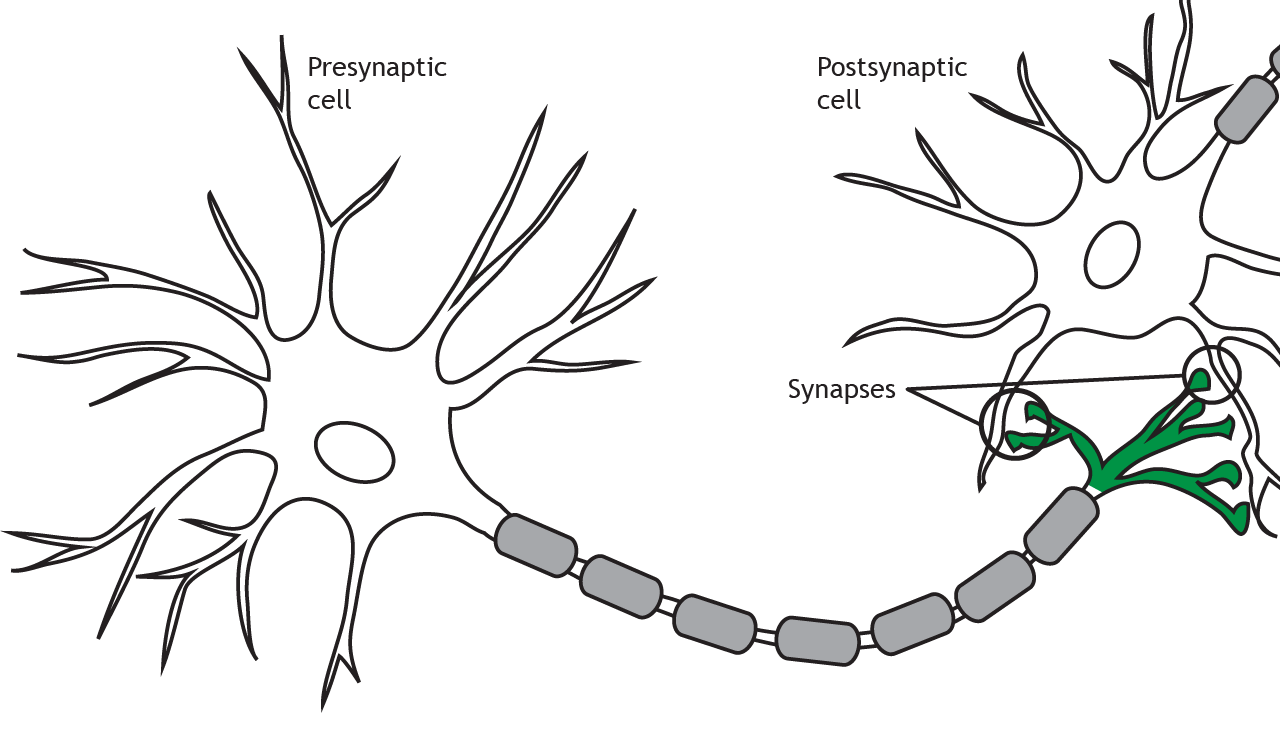
At the synapse, the terminal of a presynaptic cell comes into close contact with the cell membrane of a postsynaptic neuron.
Synapse Types
There are two types of synapses: electrical and chemical.
Electrical
Electrical synapses outnumber chemical synapses in the developing nervous system
Electrical synapses are a physical connection between two neurons. Cell membrane proteins called connexons form gap junctions between the neurons. The gap junctions form pores that allow ions to flow between neurons, so as an action potential propagates in the presynaptic neuron, the influx of sodium can move directly into the postsynaptic neuron and depolarize the cell. The response in the postsynaptic cell is almost immediate, with little to no delay between signaling in the pre- and postsynaptic neurons. Electrical synapses play an important role in the development of the nervous system but are also present throughout the developed nervous system, although in much smaller numbers that chemical synapses.
Animation 8.1. Membrane-bound proteins called connexons form gap junctions between presynaptic and postsynaptic neurons. This allows for direct exchange of ions between neurons. An action potential in the presynaptic neuron will cause an immediate depolarization of the postsynaptic membrane because the sodium ions will cross the membrane through the gap junctions. ‘Electrical Synapse – Ion Flow’ by Casey Henley is licensed under a Creative Commons Attribution Non-Commercial Share-Alike (CC BY-NC-SA) 4.0 International License. View static image of animation.
Since the gap junctions allow diffusion of ions without any obstruction, the signal can flow bidirectionally through an electrical synapse. The electrochemical gradients will drive direction of ion flow.
Animation 8.2. Since an electrical synapse is a direct, physical connection between two neurons, ions are able to flow either direction across the gap junction. ‘Bidirectional Electrical Synapse’ by Casey Henley is licensed under a Creative Commons Attribution Non-Commercial Share-Alike (CC BY-NC-SA) 4.0 International License. View static image of animation.
Additionally, small molecules like ATP or second messengers can also move through the gap junctions. These signaling molecules play an important role in cellular mechanisms, which we will see in a later chapter.
Animation 8.3. Gap junctions are large enough to allow the flow of small cellular molecules like ATP or second messengers. ‘Electrical Synapse – Small Molecules’ by Casey Henley is licensed under a Creative Commons Attribution Non-Commercial Share-Alike (CC BY-NC-SA) 4.0 International License. View static image of animation.
Chemical
Chemical synapses outnumber electrical synapses in the fully developed nervous system
Chemical synapses are the primary synapse type in the developed nervous system and do not form physical connections between the pre- and postsynaptic neurons. Instead, a space called the synaptic cleft exists between the presynaptic terminal and the postsynaptic membrane.
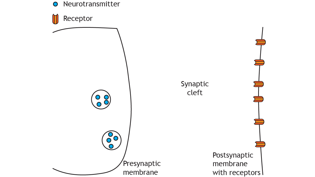
At a chemical synapse, the depolarization of an action potential reaching the presynaptic terminal causes release of neurotransmitters, which act on specialized receptors located in the cell membrane of the postsynaptic neuron. The structure and function of chemical synapses make them slower than electrical synapses and permit signaling in only one direction.
Animation 8.4. An action potential causes release of neurotransmitters from the presynaptic terminal into the synaptic cleft. The transmitters then act on neurotransmitter receptors in the postsynaptic membrane. ‘Chemical Synapse – Neurotransmitter Release’ by Casey Henley is licensed under a Creative Commons Attribution Non-Commercial Share-Alike (CC BY-NC-SA) 4.0 International License. View static image of animation.
Synapse Location
As we discuss synaptic transmission, we will focus mainly on axodendritic synapses, in which the presynaptic terminal synapses on the dendrites of the postsynaptic cell. But synapses can also be located between the terminal and the cell body of the postsynaptic cell, called axosomatic, or even between the terminal and the axon of the postsynaptic cell, called axoaxonic.

Conclusion
Both electrical and chemical synapses play vital roles in neural communication. Electrical synapses provide rapid, bidirectional signal transfer, important in development and some adult processes. Chemical synapses, being slower and unidirectional, allow for more complex signal modulation and integration, forming the predominant communication method in the developed nervous system.
Key Takeaways
- Electrical synapses allow direct ion flow between neurons through gap junctions formed by connexons, enabling rapid and bidirectional communication.
- Chemical synapses rely on neurotransmitters to bridge the synaptic cleft, facilitating slower, unidirectional signaling.
- Gap junctions in electrical synapses can also permit the passage of small molecules, like ATP and second messengers, aiding in cellular communication.
- Chemical synapses occur in diverse forms, such as axodendritic, axosomatic, and axoaxonic, depending on the postsynaptic connection site.
- The slower signaling in chemical synapses allows for greater modulation and complexity in communication compared to electrical synapses.
Test Yourself!
Try the quiz more than once to get different questions!
