
14 Reproduction
Andrea Bierema
Learning Objectives
Students will be able to:
- Define and identify examples of sexual and asexual reproduction.
- Explain how cell division is a part of reproduction.
- Describe and identify from real photos what happens to chromosomes during mitosis and meiosis.
- Define and identify sex cells.
- Define fertilization.
- Distinguish between chromatid replicates and chromosome pairs.
Note on Sex and Gender
This chapter highlights topics regarding reproduction and sex and does not refer to gender, which is a different concept.
Sex refers to a set of biological attributes in humans and animals. It is primarily associated with physical and physiological features including chromosomes, gene expression, hormone levels and function, and reproductive/sexual anatomy. Sex is usually categorized as female or male but there is variation in the biological attributes that comprise sex and how those attributes are expressed.
Gender refers to the socially constructed roles, behaviors, expressions, and identities of girls, women, boys, men, and gender-diverse people. It influences how people perceive themselves and each other, how they act and interact, and the distribution of power and resources in society. Gender identity is not confined to a binary (girl/woman, boy/man) nor is it static; it exists along a continuum and can change over time. There is considerable diversity in how individuals and groups understand, experience, and express gender through their roles, the expectations placed on them, relations with others, and the complex ways that gender is institutionalized in society.
Sexual and Asexual Reproduction
Reproduction is the production of offspring. This can happen in a variety of ways and is usually separated into sexual and asexual reproduction.
Sexual reproduction: Reproduction giving rise to offspring that have genetically unique combinations of genes (involves meiosis, a cell division process that creates sex cells).
Asexual reproduction: Reproduction that results in offspring that is genetically identical to the reproducing individual. Note that this is a different term than human asexual identity.
Exercise
Consider the definitions of sexual and asexual reproduction above as you complete the following exercise.
The rest of this section goes into more detail about the differences between sexual and asexual reproduction at the cellular level.
Cell Division
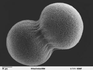
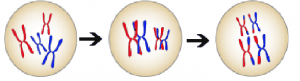
Reproduction requires cell division- either for creating sex cells (for sexual reproduction) or the reproduction itself (asexual reproduction). Cell division is when one cell divides into two- as the image below illustrates. But, what happens in the cell during cell division?
DNA Replication
As a cell prepares for division, some of the organelles duplicate- often by dividing. One of the key processes that need to happen is for the DNA to duplicate, or as it is often referred to, replicate. What would happen if the DNA did not duplicate before cell division? Recall from an earlier chapter that every DNA molecule contains genes. If all of these genes were not replicated, then only some of them would go to one cell and others to another cell and the cell would not have the complete map to create the proteins it needs to survive and function.
Chromosomes
See the chapter Gene Expression Overview to review the content on eukaryotic DNA and chromosomes. Below is a graphic that shows how DNA is packaged into a chromosome.
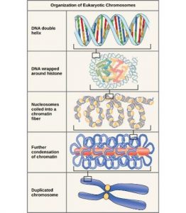
After DNA replicates, it condenses into visible (under a microscope, that is) chromosomes. The original molecule and its replicate bind together forming one chromosome that is composed of two chromatids.
Exercise
Complete the exercise below based on this fact: After DNA replicates, the chromatids of the original and new DNA molecules bind together.
Note: The term “chromosome” is used to describe two chromatids (i.e., two DNA molecules) bound together or a single chromatid (i.e., one DNA molecule).
Types of Cell Division
After the DNA is replicated, the cell finishes preparing for division (e.g., proteins needed for division are synthesized). Cell division results in new cells. During binary fission and mitosis, it results in two identical cells that are also identical to the parent cells. Meiosis, on the other hand, results in four unique cells.
Binary Fission and Mitosis
Binary fission is a form of reproduction that produces two unicellular offspring that are identical to each other and the parent cell. When the parent cell has a nucleus (and therefore, cell division includes the division of a nucleus), it is called mitosis.
The rest of this section will focus on eukaryotic cells (i.e., cells with a nucleus and more than one chromosome), which divide via mitosis. Mitosis occurs for three main reasons:
- Asexual reproduction: creates two identical, unicellular offspring
- Growth: a multicellular organism produces more cells as it grows
- Repair: old cells are replaced via the division of live cells
The main processes that occur in mitosis are that the chromosomes (those with two identical chromatids- the original and replicate DNA molecules) line up in the middle of the cell and the chromatids are pulled apart from one another by spindles and move to opposite sides of the cell. Once this happens, the same DNA exists in both halves of the cell and the cell can divide into two.
The video below illustrates what this separation of paired chromatids looks like.
The next video (Inoue, Bajer, and Mole-Bajer, 2011) illustrates what happens to the chromatids; the spindle fibers are not visible. The video begins right after the DNA condenses into chromatids after DNA replication. It illustrates the chromatids continuing to condense and gradually travel to the center of the cell. Once in the center (halfway through the video), it almost looks like the chromosomes duplicate, but it is actually just the chromatids separating from one another. Once they separate, a cell membrane begins to form in the middle and the chromatids become less condensed and, therefore, more difficult to see. A nuclear envelope/membrane forms around the genetic material on each side.
Exercise
Complete the following activity after viewing the videos above.
Meiosis
Unlike binary fission and mitosis, meiosis includes two rounds of cell division and produces four, unique cells. This process creates sex cells (or in the case of plants, the precursors of sex cells).
There are two main events that occur during meiosis:
- The separation of chromosomes that have similar genetic information.
- The separation of identical chromatids (similar to mitosis).
Most organisms that reproduce sexually have two sets of every gene- albeit, different variations of these genes. This is because they receive genetic material from their parents’ egg and sperm cells. Because genes are located on chromosomes, the chromosomes of these offspring can be paired according to the genes located on them.
Before meiosis begins, the chromosomes with the same genes pair up. These chromosome pairs will be separated into different cells during the first cell division of meiosis.
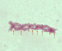
Under the microscope, the separation of the chromosome pairs looks similar to separating identical chromatids. During this first division of meiosis, rather than chromosomes forming a single line that then divides into separate chromatids, the chromosome pairs line up and the pairs are separated from each other.
The video (Oldenbourg and LaFountain , 2010) below begins with the chromosome pairs lined up in the middle of the cell and then they are separated by spindle fibers. These cells are from an insect spermatocyte.
Exercise
Complete the following activity after reading the above information and watching the video.
Chromosome Recombination Increases Genetic Diversity
Before meiosis officially begins, not only does the DNA replicate, creating chromatid replicates that bind together, recombination occurs. Recombination introduces new gene combinations into populations by “crossing” over chromosomes of the same pair. That is, once the chromosomes find their matched pairs, they exchange some DNA with each other. The exchange is variants of the same genes- so offspring still receive the full instructions to produce it. Because of this shuffling, genes from the parent’s female parent and genes from the parent’s male parent can wind up next to one another on the same stretch of DNA. This is why meiosis produces four unique cells- not two sets of two unique cells.
Sex Cells
Most sexually reproducing species have two sex cell types that differ in size: egg and sperm. Eggs and sperm for each species are identified by the size of the sex cell rather than the characteristics of the individual possessing the sex cells: egg cells are larger than sperm cells. The difference in size is due to how the cytoplasm- the material in the cell- in a cell divides. For sperm cells, during meiosis, the cytoplasm divides evenly across the four cells. For egg cells, most of the cytoplasm goes into one cell; the other three that are produced via meiosis cannot be fertilized.
Some individuals only produce one type of sex cell (identified as males or females) while others may produce both sperm and eggs. Those that produce both types are called intersex (sometimes referred to as “hermaphrodites” in non-human organisms)- either sequential (they can switch from producing one sex to producing another sex cell) or simultaneous (they can produce both sex cells at one time).
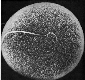
Fertilization and Early Embryonic Development
In sexually reproducing species, two sex cells- that are two different types, such as sperm and egg- combine to produce a single cell (called a zygote) with a nucleus.
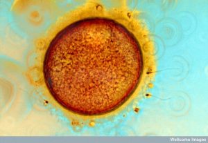
In most animals and other multicellular organisms, fertilization is followed by mitosis for growth. The following video (Inoue, Sardet, and Speksnijder, 2011) illustrates fertilization and early embryonic development.
Exercise
Take this quiz to test your understanding!
Attributions
This chapter is a modified derivative of the following sources:
Canadian Institutes of Health Research. (2020). What is gender? What is sex? This text is a modified derivative of the version available on the CIHR website.
Understanding Evolution. University of California Museum of Paleontology. “Sex and genetic shuffling: The details.” 10 August 2021 http://evolution.berkeley.edu/evolibrary/news/060101_batsars
Webster, A., Zemenick, A., & Jones, S. (n.d.). Inclusive and accurate approaches for teaching sex and gender in biology. From Biodiversify https://projectbiodiversify.org/definitions/
Videos
Rudolf Oldenbourg, James R. LaFountain (2010) CIL:9064, Nephrotoma suturalis, spermatocyte. CIL. Dataset. https://doi.org/doi:10.7295/W9CIL9064 Public domain.
Shinya Inoue, A.S. Bajer, J. Mole-Bajer (2011) CIL:11952, Haemanthus katharinae, endosperm. CIL. Dataset. https://doi.org/doi:10.7295/W9CIL11952 CC BY-NC-SA.
Shinya Inoue, Lionel Jaffe, Christian Sardet, Johanna Speksnijder (2011) CIL:11962, Phallusia mammilata, egg. CIL. Dataset. https://doi.org/doi:10.7295/W9CIL11962 CC BY-NC-SA.
Thomas Maresca, Edward Salmon (2011) CIL:26271, Drosophila melanogaster. CIL. Dataset. https://doi.org/doi:10.7295/W9CIL26271 Public domain
