
Radioactivity, Nuclear Physics and its Medical Applications
115 Nuclear Radioactivity
Learning Objectives
- Explain nuclear radiation.
- Distinguish among alpha, beta, and gamma radiation.
- Describe how ionizing radiation interacts with matter.
- Define the range of radiation and explain the factors that affect it.
The discovery of radioactivity marked the beginning of nuclear physics and fundamentally changed our understanding of matter. Scientists found that certain atomic nuclei spontaneously emit energetic particles and electromagnetic radiation without any external source of energy. These discoveries not only revealed the structure of the atomic nucleus but also led to practical applications ranging from scientific research to medicine, industry, and energy production.
Today, nuclear radiation plays an essential role in healthcare. Radioactive materials are used to diagnose disease through medical imaging, treat cancer with targeted radiation therapy, sterilize medical equipment, and support biomedical research. At the same time, understanding the properties of radiation is essential for ensuring the safety of patients, healthcare professionals, and the public.
In this section, we introduce the basic properties of nuclear radioactivity, examine the different types of nuclear radiation, and explore how radiation interacts with matter. These concepts provide the foundation for understanding the medical applications of nuclear physics presented in later chapters.

Discovery of Nuclear Radioactivity
In 1896, the French physicist Antoine Henri Becquerel accidentally discovered that a uranium-rich mineral called pitchblende emitted invisible rays capable of darkening a photographic plate, even when the plate was wrapped in opaque material. These rays carried energy and were emitted continuously without any apparent external energy source. Although this initially seemed to violate the law of conservation of energy, we now understand that the energy comes from the conversion of a small amount of nuclear mass into energy, as described by Einstein's famous equation
Further investigations showed that these mysterious rays originated from the nuclei of atoms rather than from their electrons. The spontaneous emission of energetic particles or electromagnetic radiation from an unstable nucleus is called radioactivity (or nuclear radioactivity). The emitted particles or photons are collectively known as nuclear radiation. When an unstable nucleus loses energy by emitting radiation, it is said to decay, and any material containing such unstable nuclei is described as radioactive.
Several observations demonstrated that radioactivity is a nuclear phenomenon rather than an atomic one. First, the amount of radiation emitted depends only on the element itself. Uranium, for example, remains radioactive whether it is a pure metal or part of a chemical compound, and its radioactivity is essentially unaffected by temperature, pressure, or the atom's ionization state. Since these conditions influence only the electrons surrounding the nucleus, they cannot be responsible for the emitted radiation.
A second piece of evidence is the enormous energy released during radioactive decay. Typical atomic processes, such as chemical reactions or electron transitions that produce visible light, involve energies of only a few electron volts. In contrast, nuclear decay typically releases energies on the order of
or millions of electron volts per decay. These energies are far greater than those associated with ordinary atomic processes, demonstrating that the energy must originate within the atomic nucleus.
Becquerel's discovery inspired further investigations by Marie Curie and Pierre Curie. Beginning in 1898, they studied radioactive minerals and discovered two previously unknown elements: polonium, named after Marie Curie's native Poland, and radium, named for its intense radiation. These elements were far more radioactive than uranium, with radium exhibiting roughly two million times the radioactivity per unit mass.
The Curies carried out this work under extremely difficult conditions, processing more than a ton of uranium ore to isolate only about one gram of radium salt. At the time, the biological hazards of ionizing radiation were unknown, and prolonged exposure caused serious health effects among many early researchers. Marie Curie herself suffered radiation-related injuries and later developed leukemia, a disease believed to have resulted from years of occupational exposure.
In recognition of their groundbreaking work, Becquerel and the Curies shared the 1903 Nobel Prize in Physics. Marie Curie later received the 1911 Nobel Prize in Chemistry for the discovery of polonium and radium, becoming the first person—and still the only person—to receive Nobel Prizes in two different scientific disciplines. The Curie family's contributions continued into the next generation when their daughter, Irène Joliot-Curie, and her husband, Frédéric Joliot-Curie, were awarded the 1935 Nobel Prize in Chemistry for discovering artificial radioactivity.
Healthcare Connection
The same radioactive decay that once fascinated early physicists is now an essential tool in modern medicine. Carefully selected radioactive isotopes are used to diagnose disease through PET and SPECT imaging, destroy cancer cells during radiation therapy, sterilize medical equipment, and trace biological processes within the body. Understanding the nature of radioactivity is therefore fundamental for anyone entering the health professions.
Alpha, Beta, and Gamma Radiation
As scientists continued investigating radioactivity, they discovered that unstable nuclei do not all emit the same kind of radiation. Careful experiments showed that radioactive materials produce three distinct types of nuclear radiation, which were named alpha ([latex]\alpha[/latex]), beta ([latex]\beta[/latex]), and gamma ([latex]\gamma[/latex]). The names were assigned before the true nature of the radiation was known, much like the earlier naming of x-rays.
One of the first clues that these radiations were different came from observing how they behaved in electric and magnetic fields. Figure 115.2 shows the effect of passing the three types of radiation through a magnetic field. Gamma rays continue in a straight line, while alpha and beta particles bend in opposite directions. Because charged particles experience magnetic forces, these observations showed that alpha particles carry a positive charge, beta particles carry a negative charge, and gamma rays have no electric charge.
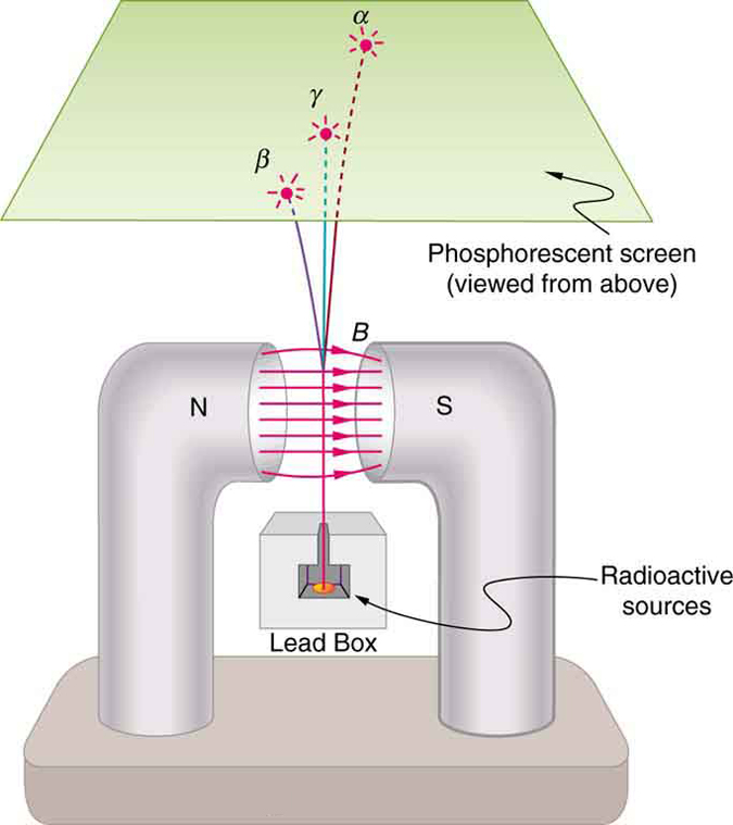
Ernest Rutherford used both electric and magnetic fields to study alpha particles in greater detail. He measured their charge to be
twice the magnitude of the electron's charge. He also found that their charge-to-mass ratio was thousands of times smaller than that of an electron, indicating that alpha particles are much more massive. By collecting alpha particles and allowing them to capture electrons, Rutherford produced helium gas and observed its characteristic spectrum. These experiments demonstrated that alpha radiation consists of helium nuclei, each containing two protons and two neutrons.
Researchers also established that beta particles have the same mass and charge-to-mass ratio as electrons. By 1902, they concluded that beta radiation consists of electrons emitted from the nucleus. These electrons are not ordinary orbital electrons that are knocked free from the atom. Instead, they are created within the nucleus during the radioactive decay process and emitted at the instant the nucleus transforms into a more stable configuration.
Gamma rays behave differently from both alpha and beta radiation because they carry no electric charge. Since they are not deflected by electric or magnetic fields, scientists proposed that they might be a form of electromagnetic radiation. In 1914, Rutherford and his collaborators confirmed this idea by observing diffraction of gamma rays from a crystal. They demonstrated that gamma radiation consists of high-energy photons emitted by an atomic nucleus.
Gamma rays are produced when an excited nucleus loses excess energy after a nuclear reaction or radioactive decay. This process is analogous to the emission of x-rays from excited atoms. The distinction between the two lies only in their origin: gamma rays originate in the nucleus, whereas x-rays originate from electron transitions outside the nucleus. At the same photon energy, gamma rays and x-rays are physically identical.
Healthcare Connection
The different types of nuclear radiation have distinct medical applications. Alpha-emitting isotopes are increasingly used in targeted cancer therapies because their short range allows them to destroy nearby cancer cells while minimizing damage to surrounding healthy tissue. Beta emitters are commonly used in therapeutic procedures and some imaging techniques, whereas gamma rays are widely used in nuclear medicine imaging because they can escape the body and be detected by external cameras.
| Type of Radiation | Typical Penetration (Range) |
|---|---|
| [latex]\alpha[/latex] particles | Stopped by a sheet of paper, a few centimeters of air, or fractions of a millimeter of biological tissue. |
| [latex]\beta[/latex] particles | Stopped by a thin sheet of aluminum or several millimeters to centimeters of tissue, depending on their energy. |
| [latex]\gamma[/latex] rays | Require several centimeters of lead or many meters of concrete for substantial shielding. |
Ionization and Range
One of the most important properties of alpha, beta, and gamma radiation is that all three can ionize matter. At the same time, they differ greatly in how deeply they penetrate materials. This penetration distance is known as the range of the radiation. Understanding ionization and range is essential because these properties determine both the biological effects of radiation and the methods used to detect and shield it.
Like x-rays, alpha particles, beta particles, and gamma rays carry enough energy to remove electrons from atoms and molecules, creating ions. The energy released during radioactive decay typically ranges from a few kiloelectron volts ([latex]\text{keV}[/latex]) to more than ten megaelectron volts ([latex]10\ \text{MeV}[/latex]), whereas only a few electron volts are required to ionize most atoms. Because of this large energy difference, even a single radioactive decay event can produce many ionizations.
Ionization is responsible for nearly all of the important effects of radiation. In living tissue, it can damage DNA and other cellular structures. In medicine, this damage is carefully controlled to destroy cancer cells during radiation therapy. Ionization is also used to detect radiation. Many radiation detectors operate by measuring the ions created when radiation passes through a gas, allowing the amount of radiation to be measured accurately.
More generally, ionizing radiation is defined as any type of radiation that has sufficient energy to remove electrons from atoms or molecules, regardless of whether the radiation originates from the atomic nucleus. Thus, x-rays and ultraviolet radiation can also be ionizing under appropriate conditions, even though they are not produced by radioactive decay.

The range of radiation is the distance radiation can travel through a material before losing essentially all of its energy. The range depends on three primary factors:
- the energy of the radiation,
- the material through which it travels, and
- the type of radiation.
Higher-energy radiation generally travels farther because it has more energy available to produce ionization. Each time radiation ionizes an atom or molecule, it loses a small amount of its energy. Consequently, particles or photons with greater initial energy are capable of producing more ionizations before coming to rest.
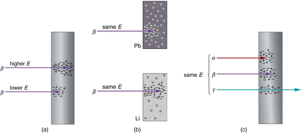
The material through which radiation travels also plays an important role. Radiation transfers energy primarily through interactions with electrons in the material. Dense materials contain more electrons per unit volume, increasing the probability of these interactions. As a result, materials such as lead are excellent radiation shields, whereas air and low-density plastics provide much less protection.
Physics Connection: Collisions
When radiation interacts with matter, it transfers energy through collisions with charged particles, primarily electrons. Because electrons have much smaller masses than atomic nuclei, they receive a much larger fraction of the transferred energy during a collision. This principle follows directly from the conservation of energy and momentum, topics introduced earlier in this text.
The type of radiation also strongly influences its range. Alpha particles carry a charge of [latex]+2e[/latex], beta particles carry a charge of [latex]-e[/latex], and gamma rays are electrically neutral. Charged particles interact much more strongly with the electrons in matter than uncharged photons. Because alpha particles have twice the electric charge of beta particles, they lose energy more rapidly and therefore travel much shorter distances.
Particle speed is another important factor. Alpha particles move at only about 5%–10% of the speed of light, whereas beta particles typically travel at relativistic speeds, and gamma rays always travel at the speed of light. Faster particles spend less time near any individual atom, reducing the probability of interaction. Consequently, beta particles generally penetrate farther than alpha particles of similar energy, while gamma rays have the greatest penetrating ability.
Healthcare Connection
The different ranges of alpha, beta, and gamma radiation determine both their medical applications and the precautions needed to use them safely. Alpha particles cannot penetrate the outer layer of skin but are highly damaging if inhaled or swallowed. Beta particles penetrate farther and require lightweight shielding such as plastic or aluminum. Gamma rays can pass completely through the human body, making them useful for diagnostic imaging but also requiring dense shielding, such as lead or concrete, to protect patients and healthcare workers.
The type of radiation also affects how far it can travel through matter, even when different types of radiation have the same energy and pass through the same material. In general, alpha particles have the shortest range, beta particles penetrate farther, and gamma rays travel the greatest distances. These differences arise primarily from the radiation's electric charge and speed.
As radiation passes through matter, it loses energy by ionizing atoms and molecules. Because each ionization requires approximately the same amount of energy, radiation that produces ionization more readily loses its energy more rapidly and therefore has a shorter range.
Charge strongly influences the likelihood of these interactions. Alpha particles carry a charge of [latex]+2q_e[/latex], beta particles carry a charge of [latex]-q_e[/latex], and gamma rays are electrically neutral. Because alpha particles have twice the magnitude of charge as beta particles, they exert stronger electric forces on nearby electrons and produce ionization more efficiently. Consequently, alpha particles lose energy quickly and travel only short distances. Gamma rays, although electrically neutral, can still interact with matter because they are high-energy electromagnetic waves. However, these interactions occur much less frequently, allowing gamma rays to penetrate much farther.
The relationship between force and momentum transfer helps explain this behavior. The change in momentum transferred during an interaction is given by
where [latex]F[/latex] is the interaction force and [latex]\Delta t[/latex] is the time over which the force acts. Radiation with smaller electric charge exerts weaker forces and therefore transfers less momentum and energy during each interaction.
Speed also plays an important role. Faster particles spend less time near atoms and molecules, reducing the probability of interaction. Alpha particles typically travel at only about 5%–10% of the speed of light, whereas beta particles usually travel at relativistic speeds. Gamma rays, being photons, always travel at the speed of light. As a result, beta particles interact less frequently than alpha particles, and gamma rays interact even less frequently.
For alpha and beta particles, the kinetic energy is given (classically) by
Because a beta particle has a mass thousands of times smaller than that of an alpha particle, it must travel much faster to have the same kinetic energy. This higher speed contributes to its greater penetrating ability.
These differences in penetration have important practical consequences. Alpha particles typically travel less than a millimeter in biological tissue and usually cannot penetrate the outer dead layer of human skin. Although they can travel several centimeters through air, simply increasing the distance from an alpha source is often sufficient for protection. However, alpha-emitting materials become extremely hazardous if inhaled, ingested, or introduced into the body through a wound, because they deposit nearly all of their energy into a very small volume of living tissue.
Beta particles can penetrate several millimeters of tissue and approximately a meter of air, making them an external radiation hazard as well as an internal one. Fortunately, relatively thin layers of shielding, such as aluminum or a few millimeters of lead, are usually sufficient to absorb most beta radiation.
Gamma rays are far more penetrating than either alpha or beta radiation. Unlike charged particles, gamma rays are gradually absorbed rather than stopped at a well-defined distance. For example, if one lead shield absorbs 90% of the incoming gamma rays, a second identical shield absorbs 90% of the remaining 10%, not 90% of the original beam. Consequently, gamma rays do not have a single, well-defined range. Instead, their intensity decreases exponentially as they pass through matter. Effective shielding therefore requires relatively thick layers of dense materials such as lead or concrete.
Healthcare Connection
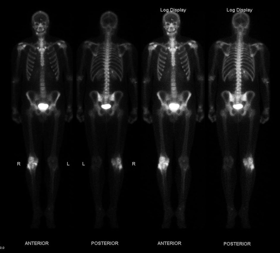
The differing ranges of alpha, beta, and gamma radiation determine their medical uses. Alpha emitters are valuable for highly targeted cancer treatments because they deliver intense damage over extremely short distances. Beta emitters are commonly used in therapeutic procedures and some diagnostic applications. Gamma rays, with their much greater penetrating power, are ideal for nuclear imaging techniques such as SPECT because they can leave the patient's body and be detected by external imaging systems.
Interactive Exploration: Beta Decay
Beta decay is a form of radioactive decay in which an unstable nucleus transforms into a more stable nucleus by emitting a beta particle. Although the decay of any individual nucleus is fundamentally random, large collections of radioactive nuclei exhibit highly predictable statistical behavior.
Use the simulation below to investigate both individual radioactive nuclei and large samples. Observe how single nuclei decay unpredictably, while the overall number of undecayed nuclei decreases in a regular pattern that can be described by a half-life.
Guided Exploration
As you explore the simulation, consider the following questions:
- Observe a single radioactive nucleus. Can you predict exactly when it will undergo beta decay?
- Switch to a collection of radioactive nuclei. How does the number of undecayed nuclei change as time passes?
- Repeat the simulation several times. Does an individual nucleus always decay at the same moment? Does the overall behavior of a large sample remain similar from one trial to the next?
- Compare the decay curves for small and large samples. Which produces a smoother curve? Why?
- Identify the particle emitted during beta decay. How does the composition of the nucleus change after the decay?
- Based on your observations, explain why radioactive decay is considered random for individual nuclei but predictable for large populations.
After completing the activity, compare your observations with the concepts discussed in this chapter. One of the defining features of radioactivity is that the exact decay time of an individual nucleus cannot be predicted, yet a sufficiently large collection of identical nuclei follows a predictable exponential decay characterized by its half-life.
Section Summary
- Some atomic nuclei are unstable and spontaneously transform into more stable nuclei by emitting energetic particles or electromagnetic radiation. This process is called radioactivity or radioactive decay.
- The three principal types of nuclear radiation are alpha particles, beta particles, and gamma rays. Alpha particles are helium nuclei, beta particles are electrons produced during nuclear decay, and gamma rays are high-energy photons emitted by excited nuclei.
- Nuclear radiation is a form of ionizing radiation because each decay releases enough energy to remove electrons from atoms and molecules.
- The range of radiation depends on its energy, the material through which it travels, and the type of radiation. Alpha particles have the shortest range, beta particles penetrate farther, and gamma rays are the most penetrating.
- Radiation detectors and dosimeters operate by measuring the ionization produced as radiation passes through matter, making ionization the basis for both radiation detection and many biological effects of radiation.
Conceptual Questions
- Suppose the range for [latex]5.0\ \text{MeV}\ \alpha[/latex] radiation is known to be 2.0 mm in a certain material. Does this mean that every [latex]5.0\ \text{MeV}\ \alpha[/latex] particle that enters the material travels exactly 2.0 mm, or is the quoted range an average value with statistical variations in the distances traveled? Explain.
- What is the difference between [latex]\gamma[/latex] rays and characteristic x-rays? Is either type of radiation necessarily more energetic than the other? Which can have the highest energies?
- Ionizing radiation interacts with matter by scattering from electrons and atomic nuclei. Based on the conservation of energy and momentum, explain why electrons generally absorb more energy than nuclei during these interactions.
- What observations indicate that radioactivity originates in the atomic nucleus rather than in the electrons surrounding the nucleus?
- What is the source of the energy emitted during radioactive decay? Identify the conservation law that was modified to account for this energy source and explain how it was extended.
- Consider Figure 115.2. If the magnetic field were replaced by an electric field, with the positive plate in the position of the north pole and the negative plate in the position of the south pole, in which directions would the [latex]\alpha[/latex], [latex]\beta[/latex], and [latex]\gamma[/latex] radiations be deflected?
- Explain how an [latex]\alpha[/latex] particle can have a greater range in air than a [latex]\beta[/latex] particle of the same energy traveling through lead.
- Arrange the following materials from the best radiation shield to the poorest radiation shield. Explain your reasoning in terms of how ionizing radiation loses energy in matter.
- A solid material with low density composed of low-mass atoms.
- A gas composed of high-mass atoms.
- A gas composed of low-mass atoms.
- A solid material with high density composed of high-mass atoms.
- People cleaning up radioactive spills often wear white protective coveralls made of plastic or similar materials. Which types of radiation, if any, do these suits protect against? Explain your reasoning.
Glossary
- alpha rays
- One of the three principal types of nuclear radiation. Alpha radiation consists of helium nuclei emitted from unstable atomic nuclei.
- beta rays
- One of the three principal types of nuclear radiation. Beta radiation consists of high-speed electrons emitted during certain types of radioactive decay.
- gamma rays
- High-energy electromagnetic radiation emitted from an excited atomic nucleus during radioactive decay.
- ionizing radiation
- Any type of radiation, whether nuclear or non-nuclear in origin, that has enough energy to remove electrons from atoms or molecules, producing ions.
- nuclear radiation
- Radiation emitted directly from the nucleus of an atom during radioactive decay.
- radioactivity
- The spontaneous emission of particles or electromagnetic radiation from unstable atomic nuclei.
- radioactive
- Describes a substance or object containing unstable nuclei that spontaneously emit nuclear radiation.
- range of radiation
- The distance that ionizing radiation can travel through a material before losing essentially all of its energy.
One of the three principal types of nuclear radiation. Alpha radiation consists of helium nuclei emitted from unstable atomic nuclei.
One of the three principal types of nuclear radiation. Beta radiation consists of high-speed electrons emitted during certain types of radioactive decay.
High-energy electromagnetic radiation emitted from an excited atomic nucleus during radioactive decay.
Electromagnetic radiation whose individual photons carry enough energy to remove electrons from atoms or molecules.
Radiation emitted directly from the nucleus of an atom during radioactive decay.
The spontaneous emission of particles or electromagnetic radiation from unstable atomic nuclei.
Describes a substance or object containing unstable nuclei that spontaneously emit nuclear radiation.
The distance that ionizing radiation can travel through a material before losing essentially all of its energy.
