
Wave Optics
87 Extended Topic: Microscopy Enhanced by the Wave Characteristics of Light
Learning Objectives
By the end of this section, you should be able to:
- Explain how the wave nature of light limits the resolution of optical microscopes.
- Describe the difference between resolution and contrast in microscopic imaging.
- Compare the operating principles of ultraviolet, interference, phase-contrast, polarization, and confocal microscopes.
- Explain how interference and polarization can improve image contrast in transparent biological specimens.
- Describe how modern microscopy techniques are used in medicine, biology, and biomedical research.
Microscopes are among the most important scientific instruments ever developed. They have transformed medicine, biology, materials science, and physics by allowing researchers to observe structures that are far too small to be seen with the unaided eye. Many of the discoveries that underpin modern healthcare—from identifying bacteria and viruses to studying individual cells and tissues—have been made possible through advances in microscopy.
The continued development of microscopes relies heavily on physics. As our understanding of light, electromagnetic waves, and image formation has improved, scientists have developed increasingly sophisticated microscopes capable of revealing smaller structures, producing higher-contrast images, and even creating three-dimensional views of living cells. In this section, we explore how the wave properties of light both limit and enhance the performance of modern microscopes.
Resolution: The Fundamental Limit
The ability of a microscope to distinguish two closely spaced objects is called its resolution. Resolution is ultimately limited by the wave nature of light. Because light undergoes diffraction when it passes around small objects or through small openings, details much smaller than the wavelength of the light cannot be resolved clearly.
As a general rule, structures significantly smaller than one wavelength of the imaging light are difficult or impossible to distinguish using conventional optical microscopes. Visible light has wavelengths ranging from approximately [latex]380\ \text{nm}[/latex] (violet) to [latex]760\ \text{nm}[/latex] (red), while individual atoms are typically about [latex]0.1\ \text{nm}[/latex] in diameter. Consequently, ordinary light microscopes cannot directly image individual atoms.
This limitation is not unique to visible light. Every imaging system is constrained by the wavelength of the waves it uses. For example, radar can easily detect the presence and approximate size of an aircraft because radio waves have wavelengths of several centimeters or more. However, those wavelengths are far too large to reveal small details such as rivets or surface scratches.
Healthcare Connection
Resolution is a critical consideration in medical imaging. Pathologists rely on microscopes capable of distinguishing individual cells and subcellular structures, while radiologists balance wavelength and resolution when choosing imaging techniques such as ultrasound, X-ray imaging, magnetic resonance imaging (MRI), or optical coherence tomography (OCT). Selecting the appropriate wavelength allows clinicians to visualize anatomical structures at the scale required for diagnosis.
Making Connections: Waves
Every imaging technique is fundamentally limited by the wavelength of the probe being used.
- Medical ultrasound uses sound waves and cannot resolve structures much smaller than the ultrasound wavelength.
- Sonar systems are limited by the wavelength of sound traveling through water.
- Optical microscopes are limited by the wavelength of visible light.
- Electron microscopes achieve much higher resolution because electrons have extremely short de Broglie wavelengths.
Later in your study of modern physics, you will learn that these limits are connected to the wave nature of matter and to Heisenberg's uncertainty principle.
Using Shorter Wavelengths
One straightforward way to improve resolution is to image with shorter wavelengths.
Ultraviolet (UV) microscopes use ultraviolet light, whose wavelengths are shorter than those of visible light. Because diffraction decreases as wavelength decreases, UV microscopes can resolve somewhat finer details than conventional light microscopes.
However, UV microscopy also presents several practical challenges. Ultraviolet radiation can damage living tissues, ordinary glass absorbs much of the UV spectrum, and specialized lenses and detectors are required. For these reasons, UV microscopy is used primarily in specialized research applications rather than routine clinical imaging.
Even shorter wavelengths—such as X-rays—or particles with wave-like properties, such as electrons, can achieve dramatically higher resolution. These imaging techniques will be discussed in later chapters.
The Importance of Contrast
High resolution alone does not guarantee a useful image. Many biological specimens are nearly transparent and absorb very little visible light, making them difficult to distinguish from their surroundings.
Contrast is the difference in brightness or intensity between an object and its background. Without sufficient contrast, important cellular structures may be invisible even if the microscope has excellent resolution.
Biologists and healthcare professionals often improve contrast by staining tissues with dyes or fluorescent molecules that selectively bind to particular cells or cellular components. Although highly effective, staining usually requires specimen preparation and may not be suitable for observing living cells.
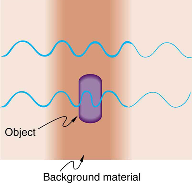
Fortunately, physics provides another solution. Because light behaves as a wave, differences in the optical path length through different regions of a specimen produce changes in phase. These phase differences can be converted into differences in intensity through interference, greatly enhancing contrast without necessarily requiring stains.
Figure 87.1 illustrates this principle. As light passes through regions with different refractive indices, its wavelength and phase change slightly. When the emerging waves are recombined, constructive and destructive interference produce brightness variations that reveal structures that would otherwise be difficult to observe.
Interference Microscopy
One method of improving image contrast without staining a specimen is interference microscopy. Instead of relying on differences in light absorption, this technique converts tiny phase differences into measurable differences in brightness.
The microscope divides light from a single source into two separate beams:
- an object beam, which passes through the specimen, and
- a reference beam, which travels through an identical optical path but does not pass through the specimen.
As the object beam travels through the specimen, variations in refractive index and thickness produce small phase shifts. When the object and reference beams are recombined, they interfere with one another. Regions where the waves reinforce each other appear brighter, while regions where they partially cancel appear darker. These intensity differences create contrast that reveals structures that would otherwise be nearly invisible.

Healthcare Connection
Interference microscopy is useful for studying transparent biological specimens that require little or no staining. Living cells, thin tissue samples, microorganisms, and cultured cells can often be examined without chemical dyes, reducing damage to the specimen while allowing researchers to observe natural cellular processes.
Phase-Contrast Microscopy
A closely related technique is phase-contrast microscopy, one of the most important advances in biological imaging. Like interference microscopy, it converts phase differences into differences in image brightness. However, it accomplishes this using a simpler optical design that is easier to align and operate.
The key component is a phase plate, an optical element that selectively shifts the phase of certain light rays before they reach the image plane. Light that has passed through the specimen interferes with background light after passing through different regions of the phase plate. The resulting constructive and destructive interference creates high-contrast images of transparent specimens.
Because most living cells are nearly transparent, phase-contrast microscopy allows them to be observed without staining or other preparation that could alter or damage the cells.
The importance of this invention was recognized with the awarding of the 1953 Nobel Prize in Physics to the Dutch physicist Frits Zernike, who developed the technique.

Phase-contrast microscopy is especially useful because it allows scientists and healthcare professionals to observe living cells in their natural state.
- No dyes or stains are required.
- Cells remain alive during observation.
- Cell division, movement, and other dynamic processes can be observed in real time.
- Transparent structures that are difficult to see with a conventional bright-field microscope become clearly visible.
Healthcare Connection
Phase-contrast microscopy is widely used in microbiology, pathology, reproductive medicine, and cell biology. It allows clinicians and researchers to examine living bacteria, blood cells, sperm cells, cultured tissues, and other transparent specimens without staining, making it an indispensable tool for biomedical research and diagnostic laboratories.
Polarization Microscopy
Another technique that improves image contrast is polarization microscopy. Instead of relying on interference between light waves, this method exploits the polarization properties of light introduced in the previous section.
The microscope first illuminates the specimen with linearly polarized light. After the light passes through the specimen, it encounters a second polarizing filter, called the analyzer, whose transmission axis is oriented perpendicular to the first polarizer.
If the specimen does not alter the polarization of the light, almost no light reaches the detector, and the image appears dark. However, many biological and crystalline materials change the polarization of light by rotating its polarization direction or by exhibiting birefringence. These changes allow some light to pass through the analyzer, producing bright regions with excellent contrast.
Many polarization effects depend on wavelength, so specimens often display vivid colors that reveal differences in internal structure and composition.
Polarization microscopy is particularly useful for specimens that are:
- optically active,
- birefringent, or
- mechanically stressed.
Because different regions of a specimen can modify polarization differently, structures that appear nearly transparent under ordinary illumination become highly visible.
Healthcare Connection
Polarization microscopy is widely used in pathology and clinical laboratories. It can identify uric acid crystals in gout, calcium pyrophosphate crystals in pseudogout, collagen fibers in connective tissue, amyloid deposits, and many other biological structures that exhibit birefringence. It is also an important tool in mineralogy and materials science.
Confocal Microscopy
Many of the microscopy techniques discussed so far can be added to conventional optical microscopes. A more advanced approach is the confocal microscope, which produces high-resolution optical sections and three-dimensional images of specimens.
A conventional microscope collects light from both the focal plane and regions above and below it. This out-of-focus light reduces image sharpness, particularly when examining thick biological samples.
Confocal microscopy overcomes this problem by illuminating only a tiny region of the specimen at a time and using a small aperture, called a pinhole, to reject light originating outside the focal plane.
A laser provides an intense, monochromatic light source that excites fluorescent molecules within the specimen. The emitted or reflected light passes back through the objective lens and then through a second pinhole positioned so that only light coming from the focal plane reaches the detector. Light originating above or below the focal plane is blocked.
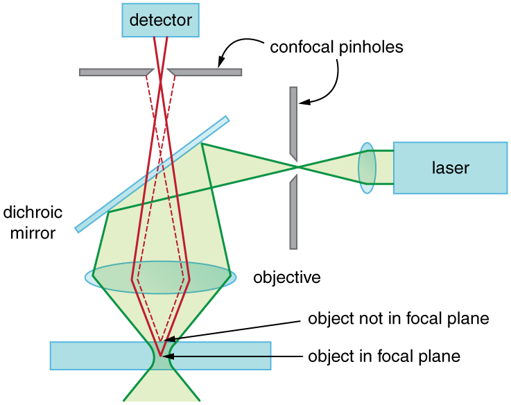
The focused laser beam is scanned across the specimen one point at a time. A computer combines these measurements into a complete image. By repeating this process at different depths, the microscope produces a series of optical slices that can be reconstructed into a detailed three-dimensional model.
Advantages of confocal microscopy include:
- Produces high-resolution three-dimensional images.
- Greatly reduces blur from out-of-focus light.
- Works well for relatively thick biological specimens.
- Allows imaging of living cells over time.
- Can visualize fluorescently labeled molecules with excellent contrast.
Healthcare Connection
Confocal microscopy has become one of the most important imaging techniques in biomedical research. It is used to study living cells, monitor cancer development, investigate infectious diseases, examine neural networks, image developing embryos, and visualize fluorescently labeled proteins inside cells. Confocal microscopes are also used clinically in ophthalmology to examine the cornea and retina at extremely high resolution.
Spectroscopic Microscopy
Modern microscopes can be combined with instruments that analyze the wavelength of the light emitted or scattered by a specimen. These techniques provide not only images but also detailed chemical information.
In one common approach, monochromatic laser light illuminates a tiny region of the specimen. Interactions between the light and the molecules in the sample slightly change the wavelength of the scattered light. Measuring these wavelength shifts reveals information about the molecular composition of the specimen.
Because every molecule produces a unique pattern of scattered wavelengths, spectroscopic techniques provide a type of molecular "fingerprint" that allows scientists to identify chemical compounds with remarkable precision.
Applications of spectroscopic microscopy include:
- Identifying chemical compounds in biological tissues.
- Monitoring biochemical reactions inside living cells.
- Studying nanomaterials and advanced materials.
- Detecting disease-related molecular changes.
- Characterizing pharmaceuticals and biomaterials.
Healthcare Connection
Techniques such as Raman microscopy and fluorescence microscopy are increasingly used in medicine to identify cancerous tissues, monitor drug delivery, analyze biopsy samples, and study cellular metabolism without requiring destructive chemical analysis.
Looking Ahead: Electron Microscopy
Even the most advanced optical microscopes remain limited by the wavelength of visible light. To observe structures much smaller than a few hundred nanometers, scientists turn to electron microscopes, which use electrons instead of visible light.
Because electrons have extremely short wavelengths, electron microscopes can resolve structures thousands of times smaller than those visible with conventional optical microscopes. They have revolutionized biology, medicine, materials science, and nanotechnology by making it possible to visualize viruses, cellular organelles, and even individual atoms.
The physics behind electron microscopy builds directly on the wave concepts introduced in this chapter and will be explored in greater detail later in the text.
Section Summary
- The resolution of an optical microscope is fundamentally limited by the wave nature of light and the effects of diffraction.
- Using shorter wavelengths, such as ultraviolet light, can improve resolution, although practical limitations restrict the widespread use of UV microscopy.
- Contrast is the difference in brightness between an object and its background. Improving contrast is often as important as improving resolution when imaging biological specimens.
- Interference microscopy enhances contrast by combining a reference beam with light that has passed through the specimen, converting phase differences into intensity differences.
- Phase-contrast microscopy uses a phase plate to transform small phase shifts into visible brightness variations, allowing transparent living cells to be observed without staining.
- Polarization microscopy improves contrast by analyzing changes in the polarization of light as it passes through birefringent or optically active materials.
- Confocal microscopy uses laser illumination, pinholes, and computer image reconstruction to reject out-of-focus light and generate high-resolution three-dimensional images.
- Spectroscopic microscopy combines microscopy with wavelength analysis to identify the chemical composition of materials using their unique optical signatures.
- Modern microscopy techniques are indispensable tools in biology, medicine, materials science, and nanotechnology.
Conceptual Questions
- Explain how wave optics allows microscopes to improve image contrast. Why is improving contrast often just as important as improving resolution when observing biological specimens?
- A beam of white light travels through water and then passes through a prism. What range of colors would you expect to emerge from the prism? Explain your reasoning based on dispersion and refraction.
Glossary
- confocal microscope
- A microscope that uses laser illumination, pinholes, and computer image reconstruction to collect light only from the focal plane, producing high-resolution optical sections and three-dimensional images.
- contrast
- The difference in brightness or intensity between an object and its background. High contrast makes structures easier to distinguish in an image.
- fluorescence microscopy
- A microscopy technique in which fluorescent molecules emit light after being excited by a laser or another light source, allowing specific structures within a specimen to be visualized.
- interference microscope
- A microscope that improves image contrast by combining an object beam with a reference beam so that differences in phase produce constructive and destructive interference.
- phase-contrast microscope
- A microscope that converts small phase differences in light passing through a transparent specimen into differences in brightness, allowing unstained living cells to be observed.
- phase plate
- An optical element used in a phase-contrast microscope to shift the phase of selected light rays, creating interference that enhances image contrast.
- polarization microscope
- A microscope that uses polarized light to reveal structures that are optically active or birefringent by analyzing changes in the polarization of light passing through a specimen.
- resolution
- The ability of an imaging system to distinguish two closely spaced objects as separate.
- spectroscopic microscopy
- A family of microscopy techniques that combine imaging with wavelength analysis to identify the chemical composition of microscopic regions within a specimen.
- ultraviolet (UV) microscope
- A microscope that uses ultraviolet light, which has shorter wavelengths than visible light, to improve image resolution. Specialized optics and detectors are required because ordinary glass absorbs much of the UV spectrum.
The ability of an imaging system to distinguish two closely spaced objects as separate.
The difference in brightness or intensity between an object and its background. High contrast makes structures easier to distinguish in an image.
A microscope that uses ultraviolet light, which has shorter wavelengths than visible light, to improve image resolution. Specialized optics and detectors are required because ordinary glass absorbs much of the UV spectrum.
A microscope that improves image contrast by combining an object beam with a reference beam so that differences in phase produce constructive and destructive interference.
A microscope that converts small phase differences in light passing through a transparent specimen into differences in brightness, allowing unstained living cells to be observed.
A microscope that uses polarized light to reveal structures that are optically active or birefringent by analyzing changes in the polarization of light passing through a specimen.
A microscope that uses laser illumination, pinholes, and computer image reconstruction to collect light only from the focal plane, producing high-resolution optical sections and three-dimensional images.
A microscopy technique in which fluorescent molecules emit light after being excited by a laser or another light source, allowing specific structures within a specimen to be visualized.
An optical element used in a phase-contrast microscope to shift the phase of selected light rays, creating interference that enhances image contrast.
A family of microscopy techniques that combine imaging with wavelength analysis to identify the chemical composition of microscopic regions within a specimen.
