
Radioactivity, Nuclear Physics and its Medical Applications
127 Radiation Dose, Biological Effects, and Radiation Safety
Learning Objectives
- Distinguish between radioactive activity, absorbed dose, equivalent dose, and effective dose.
- Convert between the common units used to measure radiation exposure.
- Explain why different types of ionizing radiation produce different biological effects.
- Describe the fundamental principles used to protect patients and healthcare workers from unnecessary radiation exposure.
Ionizing radiation is an indispensable tool in modern medicine. Every day it is used to diagnose disease, guide medical procedures, sterilize equipment, and treat cancer. At the same time, excessive exposure to ionizing radiation can damage living cells and increase the risk of adverse health effects. Understanding both the benefits and the risks of radiation is therefore essential for anyone working in healthcare.
The biological effects of radiation originate at the microscopic level. As ionizing radiation passes through tissue, it removes electrons from atoms and molecules, producing ions and highly reactive chemical species. These interactions can damage proteins, cell membranes, and, most importantly, DNA. Although cells possess remarkable repair mechanisms, severe or unrepaired DNA damage can interfere with normal cell function, trigger programmed cell death, or occasionally lead to mutations that contribute to cancer.
The previous chapter introduced the biological consequences of radiation exposure in the context of nuclear accidents and nuclear weapons. In this chapter, we focus instead on the practical aspects of radiation used in medicine: how radiation dose is measured, why different types of radiation produce different biological effects, and how healthcare professionals minimize radiation exposure while obtaining the diagnostic or therapeutic benefits of ionizing radiation.
Healthcare Connection
Radiologists, nuclear medicine physicians, radiation therapists, radiologic technologists, dentists, cardiologists, veterinarians, and many other healthcare professionals routinely work with ionizing radiation. Understanding radiation dose allows these professionals to balance diagnostic and therapeutic benefits with patient safety while following established radiation protection guidelines.
Radiation Dose: Measuring the Energy Deposited in Tissue
One of the most common misconceptions about radiation is that the strength of a radioactive source is the same as the amount of radiation received by a person. In reality, several different quantities are used to describe radiation, and each answers a different question.
Four Different Radiation Quantities
| Quantity | What It Describes | SI Unit |
|---|---|---|
| Activity | How rapidly a radioactive source decays | Becquerel (Bq) |
| Absorbed Dose | Energy deposited per kilogram of tissue | Gray (Gy) |
| Equivalent Dose | Absorbed dose adjusted for the type of radiation | Sievert (Sv) |
| Effective Dose | Equivalent dose adjusted for the sensitivity of different organs | Sievert (Sv) |
These quantities are closely related but describe different aspects of radiation exposure. A highly radioactive source does not necessarily deliver a large dose to a patient, and two patients receiving the same absorbed dose may experience different biological effects depending on the type of radiation and the tissues exposed.
Absorbed Dose
The most fundamental measure of radiation exposure is the absorbed dose, defined as the amount of ionizing energy deposited in a unit mass of tissue.
[latex]\text{Absorbed Dose}=\frac{\text{Energy Deposited}}{\text{Mass}}[/latex]
The SI unit of absorbed dose is the gray (Gy).
[latex]1~\mathrm{Gy}=1~\mathrm{J/kg}[/latex]
Historically, radiation dose was measured using the rad, where
[latex]1~\mathrm{Gy}=100~\mathrm{rad}[/latex]
Although the gray is now the standard SI unit, the rad may still appear in older publications or technical documents.
Example
If 0.50 J of ionizing energy is deposited uniformly in 2.0 kg of tissue, the absorbed dose is
[latex]D=\frac{0.50~\mathrm{J}}{2.0~\mathrm{kg}}=0.25~\mathrm{Gy}[/latex]
This quantity describes only how much energy was absorbed. It does not indicate how biologically damaging the radiation is.
Why Absorbed Dose Is Not Enough
Suppose two patients each receive an absorbed dose of 1 Gy. One is exposed to x-rays during radiation therapy, while the other receives the same absorbed dose from alpha particles inside the lungs after inhaling radioactive material.
Although both patients receive the same amount of deposited energy, the biological effects are very different. Alpha particles deposit their energy over extremely short distances, producing dense ionization tracks that cause complex DNA damage. X-rays, in contrast, deposit their energy more sparsely throughout the tissue.
For this reason, absorbed dose alone cannot fully describe the biological effects of radiation. We must also account for the type of radiation responsible for the dose.
Equivalent Dose: Accounting for the Type of Radiation
Different types of ionizing radiation do not produce the same amount of biological damage, even when they deposit the same amount of energy. For example, alpha particles lose their energy over a very short distance, producing dense tracks of ionization that are difficult for cells to repair. X-rays and gamma rays, on the other hand, spread their energy over much longer distances, producing less concentrated damage.
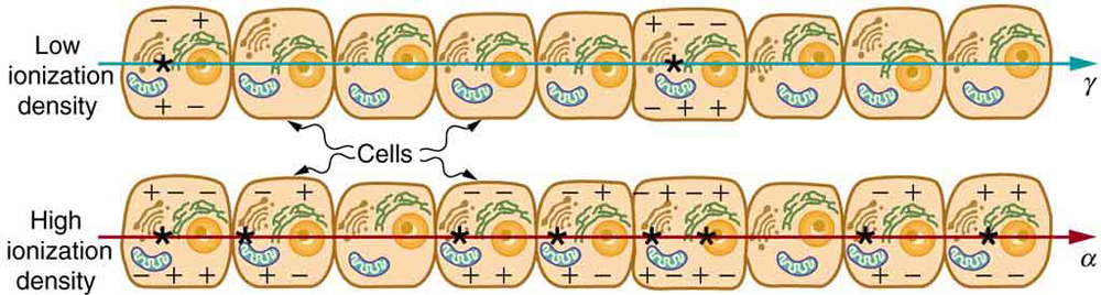
To account for these differences, radiation protection uses the concept of equivalent dose. The equivalent dose combines the absorbed dose with a radiation weighting factor, sometimes called the relative biological effectiveness (RBE) or quality factor (QF), which reflects how damaging a particular type of radiation is to living tissue.
[latex]\text{Equivalent Dose} = \text{Absorbed Dose} \times w_R[/latex]
where wR is the radiation weighting factor.
The SI unit of equivalent dose is the sievert (Sv).
[latex]1~\mathrm{Sv} = 1~\mathrm{Gy} \times w_R[/latex]
Historically, the unit rem (roentgen equivalent man) was used instead of the sievert.
[latex]1~\mathrm{Sv} = 100~\mathrm{rem}[/latex]
Typical Radiation Weighting Factors
| Radiation Type | Typical Weighting Factor (wR) |
|---|---|
| X-rays | 1 |
| Gamma rays | 1 |
| Beta particles | 1 |
| Fast neutrons | Approximately 10 |
| Alpha particles | 20 |
For x-rays, gamma rays, and most beta particles, the weighting factor is 1. As a result, the numerical values of absorbed dose (Gy) and equivalent dose (Sv) are the same. However, for alpha particles, the equivalent dose is approximately twenty times larger than the absorbed dose because alpha particles produce much greater biological damage.
Example
A patient receives an absorbed dose of 0.10 Gy from alpha particles.
[latex]\text{Equivalent Dose} = (0.10~\mathrm{Gy})(20) = 2.0~\mathrm{Sv}[/latex]
Although only 0.10 J/kg of energy was absorbed, the biological effect is comparable to about 2 Sv of x-rays or gamma rays.
Effective Dose: Accounting for the Tissue Exposed
Equivalent dose accounts for the type of radiation but does not consider where in the body the radiation is absorbed. Different organs have different sensitivities to radiation. For example, the lungs, thyroid, bone marrow, and breast tissue are generally more sensitive to radiation-induced cancer than muscle or skin.
To compare the overall health risk from different medical procedures, radiation protection introduces the concept of effective dose. Effective dose combines the equivalent dose delivered to each organ with a tissue weighting factor that reflects the relative sensitivity of that tissue.
Although the full calculation can become quite complex, the important idea is straightforward: the same equivalent dose delivered to different organs does not necessarily produce the same overall health risk.
Healthcare Connection
Effective dose is commonly used when comparing medical imaging procedures. For example, a chest x-ray, a mammogram, and a CT scan expose different organs and different amounts of tissue. Reporting the effective dose allows healthcare professionals to estimate and compare the overall radiation risk associated with these different examinations.
It is important to remember that effective dose is a tool for estimating long-term population risk. It is not intended to predict the health outcome for an individual patient, whose risk depends on many additional factors such as age, sex, and overall health.
Biological Effects of Ionizing Radiation
The biological effects of ionizing radiation begin at the atomic level. As radiation passes through tissue, it ionizes atoms and molecules, producing charged particles and highly reactive free radicals. These species can damage important cellular components, particularly DNA.
Most DNA damage is successfully repaired by the cell's natural repair mechanisms. In fact, cells experience thousands of DNA lesions each day from normal metabolic processes, ultraviolet radiation, and naturally occurring background radiation. However, if the damage is extensive or repaired incorrectly, the affected cell may respond in one of several ways:
- Successful repair: The DNA damage is repaired, and the cell continues to function normally.
- Programmed cell death (apoptosis): If the damage is too severe, the cell destroys itself to prevent abnormal growth.
- Loss of reproductive ability: The cell remains alive but can no longer divide.
- Mutation: Occasionally, damaged DNA is repaired incorrectly, potentially leading to uncontrolled cell division and, over many years, cancer.
Whether a cell survives radiation exposure depends on many factors, including the radiation dose, the type of radiation, the rate at which the dose is delivered, and the type of tissue involved.
Healthcare Connection
Radiation therapy intentionally delivers high doses of radiation to cancer cells because rapidly dividing cells are generally more sensitive to DNA damage than most healthy tissues. Normal tissues surrounding the tumor receive lower doses and are better able to repair the damage between treatment sessions.
Which Tissues Are Most Sensitive?
Not all tissues respond to radiation in the same way. Cells that divide rapidly are generally the most radiosensitive because DNA damage is more likely to interfere with cell division before repair can occur. In contrast, cells that divide infrequently tend to be more radioresistant.
Relative Radiosensitivity of Human Tissues
| More Radiosensitive | More Radioresistant |
|---|---|
| Bone marrow | Muscle |
| Intestinal lining | Mature nerve cells |
| Skin cells | Bone |
| Hair follicles | Connective tissue |
| Reproductive cells | Most mature cartilage |
This difference in radiosensitivity explains many of the early symptoms observed after large radiation exposures. Bone marrow damage reduces the production of blood cells, increasing the risk of infection and bleeding. Damage to the digestive tract can produce nausea and vomiting, while injury to hair follicles may result in temporary hair loss.
Radiation Dose and Biological Risk
The biological response to radiation depends not only on the total dose received but also on how quickly that dose is delivered. A dose received over a few minutes generally produces greater biological effects than the same dose spread over weeks or months because cells have less time to repair radiation-induced damage.
For very large radiation doses, tissue damage appears soon after exposure and is referred to as a deterministic effect. Examples include skin burns, cataracts, and acute radiation syndrome. These effects occur only after a threshold dose has been exceeded, and their severity increases as the dose increases.
At lower doses, the primary concern is the increased probability of developing cancer many years later. These are known as stochastic effects. Unlike deterministic effects, stochastic effects have no known threshold; instead, the probability of occurrence increases with dose, although the severity of the disease does not.
Key Distinction
| Deterministic Effects | Stochastic Effects |
|---|---|
| Have a threshold dose | No known threshold |
| Severity increases with dose | Probability increases with dose |
| Appear soon after high exposures | May appear years or decades later |
| Examples: skin burns, cataracts, acute radiation syndrome | Examples: cancer and hereditary effects |
For diagnostic imaging, the radiation doses are typically far below the threshold for deterministic effects. Consequently, radiation protection in medicine focuses primarily on minimizing stochastic risk while ensuring that patients receive the diagnostic or therapeutic benefits of the procedure.
Radiation Protection Principles
Because ionizing radiation has the potential to damage living tissue, healthcare professionals follow well-established safety practices whenever radiation is used. These practices are designed to keep radiation exposures as low as reasonably achievable while still obtaining the medical information or treatment needed. This philosophy is known as the ALARA principle, which stands for As Low As Reasonably Achievable.
ALARA does not mean avoiding radiation at all costs. Instead, it recognizes that radiation often provides substantial medical benefits. The goal is to minimize unnecessary exposure while ensuring that patients receive the diagnostic or therapeutic information required for appropriate care.
Healthcare Connection
Modern imaging equipment automatically adjusts the x-ray beam based on patient size and the body part being examined. This reduces the radiation dose while maintaining sufficient image quality for diagnosis, an excellent example of the ALARA principle in everyday clinical practice.
Time
The radiation dose received is directly proportional to the amount of time spent near a radiation source. Reducing the exposure time decreases the total dose received.
This principle is particularly important for healthcare workers who routinely perform fluoroscopic procedures, prepare radiopharmaceuticals, or work near patients who have received radioactive tracers.
[latex]\text{Dose} \propto \text{Exposure Time}[/latex]
Distance
Increasing the distance from a radiation source is one of the simplest and most effective methods of reducing exposure. As radiation spreads outward from a small source, its intensity decreases rapidly with distance according to the inverse-square law.
[latex]I \propto \frac{1}{r^2}[/latex]
where I is the radiation intensity and r is the distance from the source.
Doubling the distance from a point source reduces the radiation intensity to approximately one-quarter of its original value.
Example
If a technologist receives a dose rate of 4 mSv/h while standing 1 m from a radioactive source, moving to 2 m away reduces the dose rate to approximately 1 mSv/h.
This dramatic reduction illustrates why healthcare workers step away from patients immediately after administering radiopharmaceuticals whenever possible.
Shielding
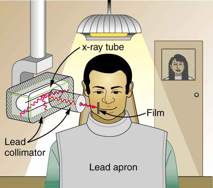
Shielding places absorbing material between the radiation source and the individual. The most effective shielding material depends on the type of radiation involved.
| Radiation Type | Typical Shielding Material |
|---|---|
| Alpha particles | Paper, clothing, or the outer layer of skin |
| Beta particles | Plastic, acrylic, or aluminum |
| X-rays and gamma rays | Lead, tungsten, or thick concrete |
| Neutrons | Water, polyethylene, or concrete containing hydrogen-rich materials |
In medical imaging departments, structural shielding is incorporated into walls, doors, windows, and equipment. Staff members may also use mobile lead barriers or syringe shields when handling radioactive materials.
Monitoring Radiation Exposure
Healthcare workers who routinely work with ionizing radiation wear personal dosimeters that continuously monitor their occupational exposure. These devices do not protect against radiation; instead, they record the accumulated dose so that exposure can be tracked over time.
Depending on the workplace, dosimeters may be worn on the chest, at the collar outside a lead apron, or on the wrist or hand when radioactive materials are handled directly. The recorded doses are reviewed regularly to ensure that occupational exposures remain well below regulatory limits.
Healthcare Connection
Radiologic technologists typically leave the examination room during routine x-ray exposures because increasing the distance from the x-ray source and standing behind a shielded control booth dramatically reduces occupational dose. In contrast, patients receive radiation only when medically indicated and usually for a very short period of time.
Special precautions are also taken for pregnant workers because the developing fetus is more sensitive to ionizing radiation than most adult tissues. Healthcare facilities may modify work assignments or provide additional monitoring to ensure that fetal radiation exposure remains well below recommended limits.
Radiation Dose in Medical Imaging
Patients are exposed to ionizing radiation during many medical imaging procedures, including x-ray radiography, computed tomography (CT), fluoroscopy, nuclear medicine, and positron emission tomography (PET). The amount of radiation received depends on the type of examination, the body part being imaged, the patient's size, and the imaging equipment used.
Radiation dose from diagnostic imaging is typically reported as effective dose, measured in millisieverts (mSv). Effective dose provides a way to compare the overall biological risk associated with different imaging procedures, even though different organs receive different amounts of radiation.
Typical Effective Doses from Medical Imaging
| Procedure | Typical Effective Dose (mSv) |
|---|---|
| Dental x-ray | 0.005 |
| Chest x-ray | 0.02 |
| Mammogram | 0.4 |
| Head CT | 2 |
| Chest CT | 7 |
| Abdomen and pelvis CT | 8–10 |
| Bone scan (Tc-99m) | 4–6 |
| PET/CT (whole body) | 15–25 |
| Average annual natural background radiation | Approximately 3 |
These values are only approximate and vary depending on the imaging protocol, patient characteristics, and equipment. Advances in detector technology and image reconstruction algorithms continue to reduce radiation doses while maintaining excellent diagnostic image quality.
Healthcare Connection
A chest CT scan typically delivers several millisieverts of radiation, which is greater than the annual dose received from natural background radiation. However, when evaluating conditions such as pulmonary embolism, severe trauma, or suspected cancer, the diagnostic information obtained from the CT scan far outweighs the small potential long-term radiation risk.
Balancing Risk and Benefit
Although ionizing radiation has the potential to damage living tissue, medical imaging is performed only when the expected clinical benefit outweighs the potential risk. For most diagnostic procedures, the radiation dose is low, and the information obtained can significantly improve patient care by allowing earlier diagnosis, more accurate treatment planning, or the avoidance of unnecessary surgery.
Radiation therapy provides an even clearer example of this balance. In radiation therapy, doses that are hundreds or even thousands of times larger than those used for diagnostic imaging are intentionally delivered to tumors in order to destroy cancer cells. Sophisticated treatment planning allows these high doses to be concentrated within the tumor while minimizing exposure to surrounding healthy tissues.
Diagnostic Imaging versus Radiation Therapy
| Diagnostic Imaging | Radiation Therapy |
|---|---|
| Uses relatively low radiation doses | Uses very high, carefully targeted radiation doses |
| Produces medical images | Destroys cancer cells |
| Goal is diagnosis | Goal is treatment |
| Typically measured in millisieverts (mSv) | Typically measured in tens of grays (Gy) |
| Minimal damage to healthy tissue | Some damage to nearby healthy tissue is unavoidable but minimized through careful treatment planning |
Modern medicine relies on the thoughtful use of ionizing radiation. By understanding how radiation dose is measured, how biological effects occur, and how exposure can be minimized through appropriate safety practices, healthcare professionals can safely use one of the most valuable diagnostic and therapeutic tools available.
Healthcare Connection: Radon—The Invisible Radiation Hazard at Home
Most people associate radiation exposure with medical imaging or nuclear accidents, but for many individuals the largest source of annual radiation exposure is actually radon gas. Radon is a naturally occurring radioactive gas produced during the decay of uranium found in rocks and soil. Because it is colorless, odorless, and chemically inert, radon can accumulate inside homes without being noticed.
When radon is inhaled, its radioactive decay products can become trapped in the lungs. Many of these decay products emit alpha particles, which deposit their energy over extremely short distances. Although alpha particles cannot penetrate the outer layer of the skin, they can cause significant damage to lung tissue when emitted inside the body.
Long-term exposure to elevated radon concentrations is the second leading cause of lung cancer after cigarette smoking and the leading cause among people who have never smoked. The risk is especially high for individuals who both smoke and are exposed to elevated radon levels.
Because radon levels vary from one location to another, many public health agencies recommend testing homes, particularly those with basements or crawl spaces. If elevated concentrations are detected, ventilation systems and sub-slab depressurization systems can significantly reduce indoor radon levels.
Common Misconceptions About Radiation
- MRI does not use ionizing radiation. It uses magnetic fields and radio waves.
- Ultrasound does not use ionizing radiation. It uses high-frequency sound waves.
- People naturally receive radiation every day. Cosmic rays, naturally occurring radioactive materials in the Earth, and even foods containing potassium-40 contribute to background radiation.
- Nuclear medicine patients may emit small amounts of radiation for a short time. Hospitals provide instructions when temporary precautions are needed.
- The goal is not zero radiation. The goal is to keep exposures As Low As Reasonably Achievable (ALARA) while obtaining important medical benefits.
Section Summary
- Radiation exposure can be described using several different quantities, including activity (Bq), absorbed dose (Gy), equivalent dose (Sv), and effective dose (Sv).
- Absorbed dose measures the amount of energy deposited in tissue, while equivalent dose accounts for the biological effectiveness of different types of radiation.
- Effective dose considers both the type of radiation and the sensitivity of the organs exposed, allowing comparisons between different medical imaging procedures.
- Ionizing radiation can damage DNA. Most damage is repaired, but high doses may produce tissue injury, while low doses primarily increase the probability of long-term effects such as cancer.
- Radiation protection is based on the ALARA principle and the three fundamental methods of reducing exposure: minimizing time, maximizing distance, and using appropriate shielding.
- Medical imaging procedures expose patients to varying radiation doses, but when properly justified, the diagnostic or therapeutic benefits greatly outweigh the potential risks.
Conceptual Questions
- Why is the absorbed dose (Gy) alone not sufficient to describe the biological effects of radiation? Why is the equivalent dose (Sv) often more useful in healthcare?
- Alpha particles cannot penetrate the outer layer of skin, yet alpha-emitting materials can be extremely dangerous if inhaled or swallowed. Explain why.
- Radon gas is the largest source of natural radiation exposure for many people. Why does long-term radon exposure primarily increase the risk of lung cancer rather than cancers in other organs?
- Two healthcare workers perform the same fluoroscopic procedure. One remains twice as far from the x-ray source as the other whenever possible. Using the inverse-square law, explain how distance affects their radiation exposure.
- Explain the ALARA principle. Why does "As Low As Reasonably Achievable" not mean avoiding all medical radiation?
- Different materials are used to shield against alpha particles, beta particles, gamma rays, and neutrons. Explain why no single shielding material is ideal for every type of radiation.
- Why are rapidly dividing tissues, such as bone marrow and the lining of the digestive tract, generally more sensitive to ionizing radiation than muscle or nerve tissue?
- Distinguish between deterministic and stochastic effects of radiation exposure. Give one example of each.
- Why do radiologic technologists typically leave the examination room or stand behind a protective barrier during an x-ray exposure while the patient remains in the room?
- A CT scan delivers a higher radiation dose than a standard chest x-ray. Under what circumstances is performing a CT scan still the appropriate medical decision?
Problems & Exercises
- What is the equivalent dose in mSv for:
- a 0.10 Gy x-ray?
- 2.5 mGy of neutron exposure to the eye (assume wR = 10)?
- 1.5 mGy of alpha-particle exposure?
- Find the absorbed dose in Gy for:
- a 10 mSv fluoroscopic x-ray examination,
- 50 mSv of skin exposure from an alpha emitter, and
- 160 mSv of beta or gamma radiation from naturally occurring 40K in the body.
- How many grays of absorbed dose are required to deliver an equivalent dose of 40 Sv using alpha radiation?
- A patient receives 60 Gy of gamma radiation during a course of radiation therapy. What is the equivalent dose in sieverts?
- One half of the gamma rays from 99mTc are absorbed by a 0.170-mm-thick sheet of lead. Assuming each additional layer absorbs half of the remaining radiation, what total thickness of lead is required so that only one in every 1000 gamma rays passes through?
- A technologist receives an occupational dose of 0.60 mSv during a month of work. If this monthly dose remained constant throughout the year, what would be the worker's annual effective dose?
- A patient receives a chest CT scan with an effective dose of 7 mSv. Approximately how many years of average natural background radiation (3 mSv/year) does this correspond to?
- A radioactive source produces a dose rate of 12 mSv/h at a distance of 1.0 m. Assuming the source behaves as a point source, what is the dose rate at:
- 2.0 m?
- 3.0 m?
Glossary
- absorbed dose
- The amount of energy deposited by ionizing radiation per unit mass of material. The SI unit is the gray (Gy).
- ALARA (As Low As Reasonably Achievable)
- A radiation safety principle that minimizes unnecessary radiation exposure while still achieving the required medical or scientific objective.
- alpha particle
- A positively charged particle consisting of two protons and two neutrons emitted during certain types of radioactive decay. Alpha particles have high ionizing power but very limited penetration.
- background radiation
- Ionizing radiation naturally present in the environment from cosmic rays, naturally occurring radioactive materials in the Earth, radon gas, and radioactive isotopes within the human body.
- becquerel (Bq)
- The SI unit of radioactive activity, equal to one radioactive decay per second.
- deterministic effect
- A biological effect of radiation that occurs only after a threshold dose has been exceeded and becomes more severe as dose increases. Examples include skin burns and cataracts.
- A device worn by radiation workers to measure and record accumulated occupational radiation exposure.
- effective dose
- A measure of radiation exposure that accounts for both the type of radiation and the varying sensitivities of different tissues. The SI unit is the sievert (Sv).
- equivalent dose
- A measure of radiation dose that accounts for the biological effectiveness of different types of radiation by applying a radiation weighting factor. The SI unit is the sievert (Sv).
- free radical
- A highly reactive atom or molecule produced by ionizing radiation that can damage cellular structures, including DNA.
- gray (Gy)
- The SI unit of absorbed dose, equal to one joule of radiation energy absorbed per kilogram of material.
- inverse-square law
- The principle stating that radiation intensity from a point source decreases in proportion to the square of the distance from the source.
- ionizing radiation
- Radiation with enough energy to remove electrons from atoms or molecules, producing ions that can damage biological tissue.
- personal protective equipment (PPE)
- Protective equipment, such as lead aprons, thyroid shields, and protective barriers, used to reduce occupational radiation exposure when appropriate.
- quality factor (QF)
- An older term, now largely replaced by the radiation weighting factor, used to account for differences in the biological effectiveness of various types of ionizing radiation.
- radiation weighting factor (wR)
- A numerical factor used to convert absorbed dose into equivalent dose by accounting for the relative biological effectiveness of different types of radiation.
- radioresistant tissue
- Tissue that is relatively less sensitive to damage from ionizing radiation, such as muscle or mature nerve tissue.
- radiosensitive tissue
- Tissue whose rapidly dividing cells are especially susceptible to radiation damage, including bone marrow and the lining of the digestive tract.
- radon
- A naturally occurring radioactive gas produced during the decay of uranium in rocks and soil. Long-term exposure to elevated indoor radon concentrations increases the risk of lung cancer.
- rem
- A traditional unit of equivalent or effective dose. One sievert equals 100 rem.
- relative biological effectiveness (RBE)
- A measure of how effective a particular type of radiation is at producing biological damage compared with a reference radiation for the same absorbed dose.
- shielding
- The use of absorbing materials between a radiation source and a person to reduce radiation exposure.
- sievert (Sv)
- The SI unit of equivalent dose and effective dose that reflects the biological effects of ionizing radiation.
- stochastic effect
- A radiation-induced effect whose probability increases with dose but whose severity does not depend on dose. Cancer is the primary example.
- time-distance-shielding
- The three fundamental principles of radiation protection: minimizing exposure time, maximizing distance from the source, and using appropriate shielding.
- computed tomography (CT)
- An imaging technique that uses x-rays acquired from multiple angles to produce detailed cross-sectional images of the body. CT typically delivers higher radiation doses than conventional radiography but provides substantially more diagnostic information.
The amount of energy deposited by ionizing radiation per unit mass of material. The SI unit is the gray (Gy).
An acronym for As Low As Reasonably Achievable, the guiding principle of radiation protection that seeks to minimize unnecessary radiation exposure while achieving the intended medical or scientific objective.
A positively charged particle consisting of two protons and two neutrons emitted during certain types of radioactive decay. Alpha particles have high ionizing power but very limited penetration.
Ionizing radiation naturally present in the environment from cosmic rays, naturally occurring radioactive materials in the Earth, radon gas, and radioactive isotopes within the human body.
The SI unit of radioactivity, equal to one radioactive decay per second.
A biological effect of radiation that occurs only after a threshold dose has been exceeded and becomes more severe as dose increases. Examples include skin burns and cataracts.
A device worn by radiation workers to measure and record accumulated occupational radiation exposure.
A measure of radiation exposure that accounts for both the type of radiation and the varying sensitivities of different tissues. The SI unit is the sievert (Sv).
A measure of radiation dose that accounts for the biological effectiveness of different types of radiation by applying a radiation weighting factor. The SI unit is the sievert (Sv).
A highly reactive atom or molecule produced by ionizing radiation that can damage cellular structures, including DNA.
The SI unit of absorbed dose, equal to one joule of radiation energy absorbed per kilogram of material.
The principle stating that radiation intensity from a point source decreases in proportion to the square of the distance from the source.
Electromagnetic radiation whose individual photons carry enough energy to remove electrons from atoms or molecules.
Protective equipment, such as lead aprons, thyroid shields, and protective barriers, used to reduce occupational radiation exposure when appropriate.
An older term, now largely replaced by the radiation weighting factor, used to account for differences in the biological effectiveness of various types of ionizing radiation.
A numerical factor used to convert absorbed dose into equivalent dose by accounting for the relative biological effectiveness of different types of radiation.
Tissue that is relatively less sensitive to damage from ionizing radiation, such as muscle or mature nerve tissue.
Tissue whose rapidly dividing cells are especially susceptible to radiation damage, including bone marrow and the lining of the digestive tract.
A naturally occurring radioactive gas produced during the decay of uranium in rocks and soil. Long-term exposure to elevated indoor radon concentrations increases the risk of lung cancer.
A traditional unit of equivalent or effective dose. One sievert equals 100 rem.
A measure of how effective a particular type of radiation is at producing biological damage compared with a reference radiation for the same absorbed dose.
The use of absorbing materials between a radiation source and a person to reduce radiation exposure.
The SI unit of equivalent dose and effective dose that reflects the biological effects of ionizing radiation.
A radiation-induced effect whose probability increases with dose but whose severity does not depend on dose. Cancer is the primary example.
The three fundamental principles of radiation protection: minimizing exposure time, maximizing distance from the source, and using appropriate shielding.
An imaging technique that uses x-rays acquired from multiple angles to produce detailed cross-sectional images of the body. CT typically delivers higher radiation doses than conventional radiography but provides substantially more diagnostic information.
