
Geometric Optics, Vision and Optical Instruments
77 Aberrations
Learning Objectives
- Describe the major types of optical aberrations and explain how they affect image quality.
- Explain how optical engineers reduce aberrations in microscopes, cameras, telescopes, and medical imaging systems.
The thin lens model introduced in earlier chapters accurately predicts image formation for many simple situations. Real optical systems, however, are more complex. Real lenses have finite thickness, are made from materials whose refractive properties depend on wavelength, and may have imperfections or curved surfaces that differ from the idealized models. As a result, real optical systems often produce images that are slightly distorted or blurred. These imperfections are known as optical aberrations.
An aberration is any departure from ideal image formation. Depending on the design of the optical system, aberrations may reduce sharpness, distort shapes, shift colors, or prevent all of the light rays from focusing at a single point. Modern microscopes, telescopes, cameras, endoscopes, and even the human eye must all contend with optical aberrations.
Engineers spend considerable effort designing lenses that minimize these effects. In many modern optical instruments, multiple lenses made from different materials are combined so that one lens compensates for the imperfections of another.
Chromatic Aberration
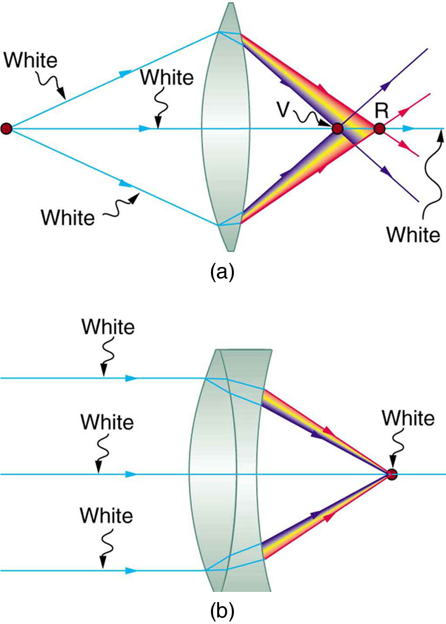
One of the most familiar aberrations is chromatic aberration. It occurs because the refractive index of glass depends on the wavelength (color) of light. Shorter wavelengths, such as violet light, experience a larger refractive index than longer wavelengths, such as red light. Consequently, different colors are bent by different amounts as they pass through a lens.
Because violet light refracts more strongly than red light, the different colors come to focus at different distances from the lens. Instead of producing a single sharp image, the lens produces slightly different images for each color, often resulting in colored fringes around high-contrast objects.
Mirrors do not suffer from chromatic aberration because reflection is independent of wavelength. This is one of the reasons that nearly all large research telescopes use mirrors instead of lenses.
Healthcare Connection
Chromatic aberration must be minimized in medical microscopes, ophthalmic instruments, and surgical imaging systems. Even small color-dependent distortions can reduce image quality when examining tissue samples, blood cells, or the retina. High-quality microscope objectives therefore contain multiple carefully designed lens elements that compensate for chromatic aberration.
Coma
Another important optical aberration is coma. This aberration occurs when light from an object located away from the optical axis does not converge to a single point after passing through a lens or reflecting from a mirror.
Instead, different regions of the optical element focus the light at slightly different locations, producing an asymmetric image that often resembles a comet with a bright head and a faint tail. This effect gives the aberration its name.
Coma is especially important in telescopes and wide-angle camera lenses because objects near the edges of the field of view are more susceptible to this distortion.
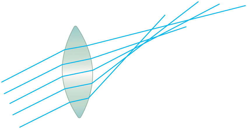 Figure 77.2: Coma occurs when light from an off-axis object fails to focus at a single point, producing a distorted, comet-like image.</caption]
Spherical Aberration
Spherical aberration arises because spherical lenses do not focus all incoming rays at exactly the same location. Rays passing through the outer portions of the lens are refracted more strongly than rays passing close to the optical axis.
As a result, peripheral rays come to focus at a different distance from the lens than central rays. Instead of producing a sharp point, the image becomes blurred.
Spherical aberration becomes more noticeable when large-aperture lenses are used because more peripheral rays enter the optical system.
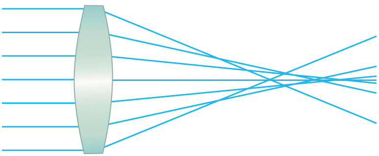 Figure 77.3: In spherical aberration, rays passing through different regions of a lens focus at different locations, reducing image sharpness.</caption]
Brightness versus Image Quality
An optical system must collect enough light to produce a bright image, but increasing the amount of light entering the system often increases aberrations as well. Consequently, optical designers must balance image brightness with image quality.
The diameter of a lens, mirror, or aperture stop determines how much light enters the system. Large apertures collect more light, making images brighter, but they also allow more peripheral rays to enter, increasing aberrations such as spherical aberration.
Reducing the aperture limits these problematic rays, often producing a noticeably sharper image. This is why squinting sometimes improves vision. By narrowing the effective aperture of the eye, only rays passing near the optical axis contribute significantly to the image, reducing blur.
Everyday Physics
If you look through a tiny hole made with your fingers or in a piece of paper, objects often appear sharper. The small opening acts like an aperture stop, blocking many of the rays responsible for optical aberrations and increasing the depth of field.
Modern Methods for Correcting Aberrations
Modern optical instruments employ a variety of techniques to reduce aberrations.
- Multiple-element lenses combine several lenses made from different materials so that one lens compensates for the aberrations produced by another.
- Aspheric lenses have carefully designed surfaces that differ from simple spherical shapes, greatly reducing spherical aberration while keeping the lens compact.
- Graded-index (GRIN) lenses have a refractive index that changes gradually throughout the material rather than remaining constant. These lenses are widely used in fiber-optic communication systems and miniature medical endoscopes.
- Adaptive optics uses computer-controlled mirrors that continuously change shape to compensate for atmospheric turbulence. This technology allows ground-based telescopes to achieve images approaching the sharpness of space telescopes.
- Computational imaging uses software to remove distortions after an image has been captured. Modern smartphone cameras, medical imaging systems, and astronomical observatories routinely combine optical corrections with sophisticated digital image processing.
Healthcare Application
Wavefront-guided LASIK surgery measures tiny optical aberrations unique to each patient’s eye before reshaping the cornea. Instead of correcting only nearsightedness or farsightedness, modern systems can also reduce higher-order aberrations, improving contrast sensitivity and night vision. Similar wavefront measurements are used in advanced ophthalmic diagnostic instruments.
Section Summary
- Optical aberrations are imperfections in image formation that prevent all light rays from focusing at a single point, reducing image sharpness or introducing distortions.
- Common aberrations include chromatic aberration, coma, spherical aberration, and astigmatism. Each arises from different properties of lenses or mirrors and affects image quality in different ways.
- Chromatic aberration occurs because the refractive index of a lens depends on the wavelength of light, causing different colors to focus at different locations.
- Coma and spherical aberration result from rays passing through different regions of an optical element, producing distorted or blurred images.
- Reducing the aperture of an optical system can improve image sharpness by limiting rays that contribute most strongly to certain aberrations, although it also decreases image brightness.
- Modern optical systems reduce aberrations using combinations of specially designed lenses, aspheric surfaces, graded-index (GRIN) materials, adaptive optics, and computational image processing.
Conceptual Questions
- Describe the major types of optical aberrations. What causes each one, and what techniques can be used to reduce or correct them?
Problems & Exercises
- Integrated Concepts.
- During laser vision correction (LASIK), a brief pulse of 193 nm ultraviolet light is projected onto the cornea. The laser produces a circular spot 1.00 mm in diameter and deposits 0.500 mJ of energy. Assuming the corneal tissue has the same thermal properties as water and is initially at 34.0°C, calculate the depth of tissue removed if the tissue is heated to 100°C and then vaporized without any additional temperature increase.
- Based on your result, discuss whether this technique allows the shape of the cornea to be controlled with high precision.
Glossary
- aberration
- An optical imperfection that prevents all light rays from converging at a single focus, resulting in image blur, distortion, or other departures from ideal image formation.
An optical imperfection that prevents all light rays from converging at a single focus, resulting in image blur, distortion, or other departures from ideal image formation.
