
1 Course Themes and Scientific Practices
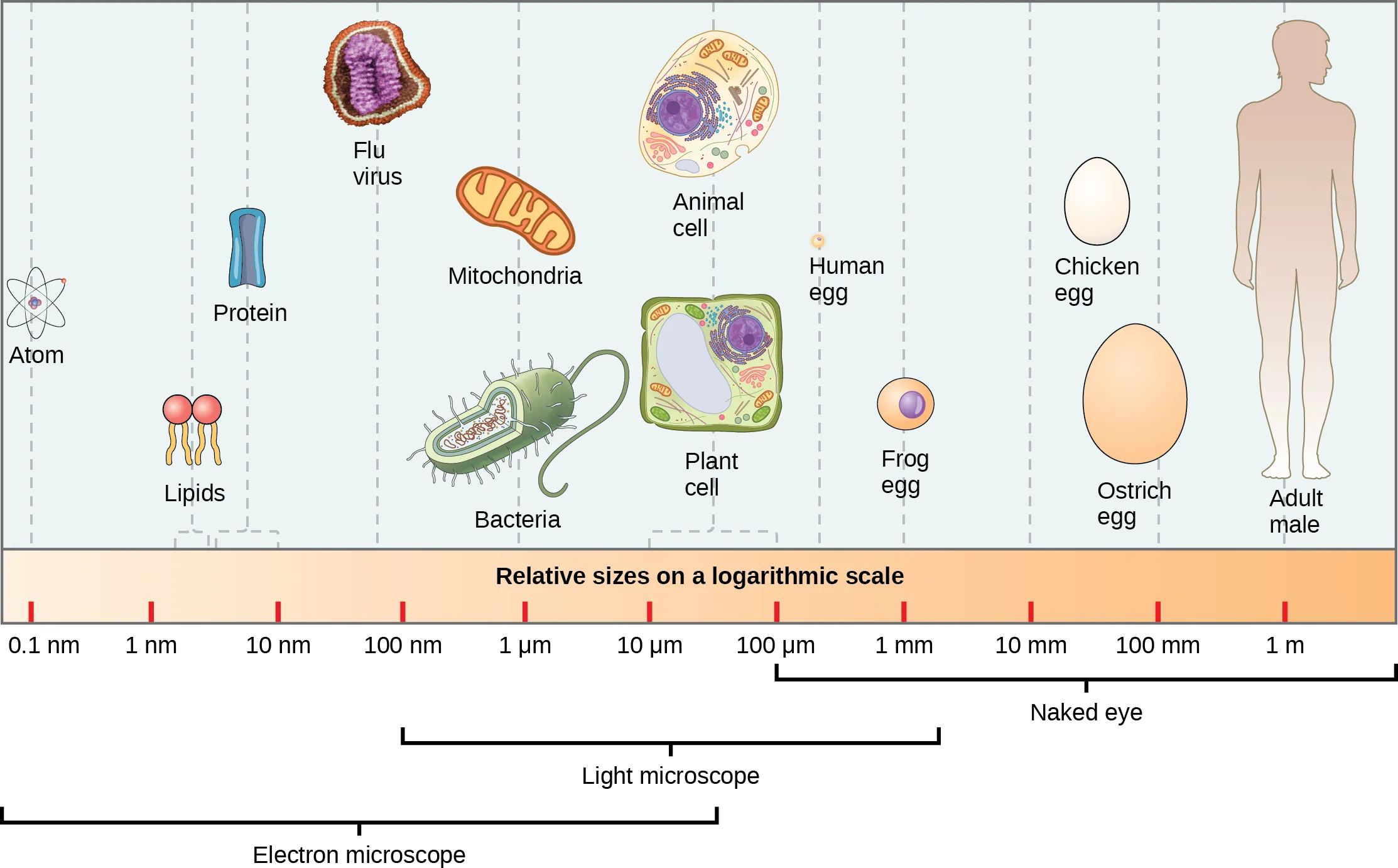
Welcome to the study of cells and molecules! Have you ever wondered why we have the traits that we have, or why things in your body work the way they do? This textbook provides the foundation (although not all the detail) for these inquiries. We will focus on the structure and behavior of cells, which are a direct consequence of the molecules in cells and their interactions with other molecules. In our exploration of how cells work, we will often be discussing molecules and processes that are not observable without the aid of microscopes or other techniques (Figure 1.1). However, by studying cells and molecules, we can better understand how organisms function, and how genetics and environmental influences affect functions and traits. These molecular and cellular insights are useful in many ways, including for understanding our health and solving medical and environmental problems.
We focus on cells as all living organisms are made of cells, and cells exhibit all the key features of life. For some organisms, such as bacteria or yeasts, the cell is the entire organism, while some multicellular organisms such as yourself contain trillions of cells. Yet even cells in a multicellular organism, while communicating and cooperating with other cells, function as individual units. Each cell has its own genetic material in the form of DNA, which contains the instructions to build RNA and proteins that are needed for cellular functions. Each cell must also be capable of obtaining materials from the environment and transforming them into other molecules or useful forms of cellular energy to carry out cellular functions. One such cellular function is creating an organized internal environment. Each cell also senses its environment and may change its behavior based on signals received. Finally, a cell must reproduce to create more cells, and all cells observed today came from existing cells.
Chapter Outline
Section 1.1 Themes in Cells and Molecules
Section 1.3 Scientific Arguments
Section 1.1 Themes in Cells and Molecules
This textbook introduces fundamental aspects of cellular structure and function, and in the exploration of each topic, we encourage you to view the content through each of four themes described below and visualized in Figure 1.2.

Theme 1: From Structure to Function
A pervasive theme throughout biology is the connections among a molecule’s shape, interactions, and its function. The shape of a molecule is chemically determined: the atoms and chemical bonds in the molecule dictate the overall structure of a molecule and its chemical properties. Those in turn determine how that molecule interacts with other molecules, or other aspects of the molecule’s function. We will see many examples of molecules that “fit together” in a way that supports a larger biological process. Conversely, we might expect that if the shape of the molecule were altered, it would no longer be able to carry out the same function in cells, and may not function at all.
For example, an enzyme, which is a type of protein, can speed up chemical reactions by first binding to the substrate molecule involved in the reaction. The enzyme’s shape and chemical properties must be complementary, or “fit” with the shape and chemical properties of the substrate molecule, to result in the products being observed.
Theme 2: Genes, Environment, and Phenotypes
As with all proteins in the cell, the shape of the enzyme and the interactions it can have are encoded in the cell’s genome and is influenced by environmental factors. The DNA sequence containing instructions for making a particular protein is the gene. Through gene expression, the information in DNA is used to make an RNA, and that RNA may be used to make a protein. Typically, the actions of many proteins, influenced by the environment, are integrated to result in a phenotype, or trait. For example, the ability of a cell to produce a certain molecule may depend on the actions of many enzymes, each encoded by its own gene that is independently expressed in the cell. Environmental factors, such as dietary molecules, can further regulate which genes are expressed or modify the actions of enzymes.
Because every protein is encoded by the cell’s DNA, a protein’s structure and potentially its function can be altered by changing the DNA sequence. Indeed, all of us are walking around with slightly different versions of many proteins due to slightly different gene sequences among humans. This genetic variation (genotypes), along with different environmental influences, gives rise to the many different observable phenotypes, or traits, in populations.
Theme 3: Energy, Matter, and Metabolism
To carry out functions in the cell—whether to build RNA and proteins, to reproduce, to respond to the environment—requires reorganization of molecules, which often requires energy. To satisfy the laws the thermodynamics, cells must couple energy-requiring processes with processes that release sufficient energy. In photosynthetic organisms, the process of photosynthesis uses the energy of light along with CO2 from the air to synthesize carbohydrates, which can then be transformed into other organic food molecules. Photosynthesis also produces O2, which is essential for cellular respiration performed by most organisms. In cellular respiration, the breakdown of food molecules is coupled to the production of adenosine triphosphate (ATP) from adenosine diphosphate (ADP) and inorganic phosphate (Pi). In turn, the breakdown of ATP into ADP and Pi is coupled to many of the energy-requiring processes in cells.
Cells require many enzymes to facilitate the reactions of metabolism at reasonable rates and under cellular conditions. These enzymes are proteins encoded by DNA, and as noted above, changes to the genetic information could change the functions of these proteins. Thus, the ability to carry out metabolic reactions to produce certain molecules is an observable phenotype that is related to an organism’s genotype. Moreover, environmental influences (think diet and exercise!) also affect one’s metabolism.
Theme 4: Regulation
Molecular processes in cells, including gene expression, metabolism, and cell division, are dynamically regulated in response to internal and environmental stimuli. For example, bacteria alter their gene expression based on available nutrients, altering their metabolism in response to their environment. In multicellular organisms, cells reproduce only in response to specific signals received from other cells, which in turn cause many changes within the receiving cell. The process of cell division requires extensive metabolic changes to accommodate new gene expression, DNA replication, and the reorganization of cellular materials to divide one cell into two.
Section 1.2 Scientific Models
One of the challenges of cell and molecular biology is that most of the molecules and structures involved are too small to see without the aid of a microscope (see Figure 1.1). To help us “visualize” the molecules in cells, we will use symbols to represent these molecules, and build models to show the relationships among them. The scientific models we create will be based on evidence and capture our understanding of how a process works and how the pieces fit together. Once built, models are tools to make predictions of how a change to one molecule or part of the process would affect other parts of the process. For a research lab, those predictions would be tested experimentally, and the results of the experiment would be used to further modify the model.
You will see models through this textbook and in all class resources, and will be building and interpreting models regularly. We encourage you to regularly draw out models to make sense of the many molecular processes that you will be learning, as they are a great way for you to also communicate your understanding so that you can get feedback from peers or teaching team! The models in Figure 1.2 summarize some of the big themes of this textbook, and you are encouraged to come back to this figure as you learn more detail about each of the processes depicted. Here are some tips about models to keep in mind as you review the models in this book and build your own:
Labels
Because the symbols in models represent things we can’t ordinarily see, they may not be intuitive to interpret. However, the labels on models will help you to understand what is being depicted, and you should carefully label molecules in the models you draw, for the same reason.
Simplifications to Emphasize the Goal
All models are simplifications, meant to emphasize certain points while limiting or leaving out certain details that could distract from the main message. A realistic cell model would contain so many molecules that are so tightly packed together, it would be hard to know what to focus on! Thus, we might show one protein or one structure when in reality, tens to thousands of each might be present in the cell. For each figure you evaluate, try to identify the main message being conveyed. Ask yourself, “What is this model trying to teach me?”
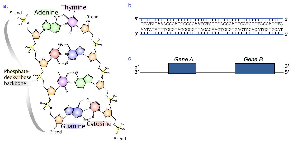
Sometimes even the same molecule will be represented in different ways, depending on the goal or emphasis of the model. For example, when discussing the chemical structure of DNA, it is important to show the atoms of a DNA strand (Figure 1.3a). However, the chemical details might be omitted in a discussion of gene expression that is focused on the genetic information—the As, Ts, Gs, and Cs of a DNA sequence (Figure 1.3b). Finally, in a “zoomed-out” version of a large DNA segment, even the sequence details will be left out to emphasize the relative location of genes (Figure 1.3c). In all cases, the labels on the models and in the caption will tell you what molecule you are looking at. We encourage you to compare multiple models of the same molecule, reflecting on what each model emphasizes and why!
Arrows
Biologists tend to use a lot of arrows in their models, but the meanings of arrows can differ depending on the context. In general chemistry, arrows usually indicate chemical reactions, and that meaning also holds true in some biological models. However, arrows more commonly indicate links between molecules in a process, the movement of a molecule, or the passage of time (for example, before -> after).
Let’s look again the model of gene expression at the top right section of Figure 1.2. Here we see DNA -> RNA. One interpretation of this model is that the DNA is a reactant and RNA is a product, with the DNA molecule turning into the RNA molecule. However, the building of an RNA molecule is somewhat like following a cookbook recipe, which gives the correct ingredients and the order in which to assemble them. The DNA sequence is the recipe, not an ingredient! Thus, the arrow in the model represents the process that transfers information from DNA to RNA, not a chemical reaction of DNA becoming RNA.
It will take time to learn the biological context and become comfortable with the different meanings of arrows. One tip is to aid your understanding is to redraw the models from memory, saying aloud or describing in writing what is happening at each step.
Section 1.3 Scientific Arguments
Drawing models is just one of the approaches we can use to make sense of biological information. Another key practice is argumentation, which is using evidence and reasoning to make a claim, or conclusion, about a biological phenomenon. You are undoubtedly familiar with the practice of argumentation in other contexts and likely have engaged in argumentation yourself, if you’ve ever been persuaded or tried to persuade someone to a particular viewpoint. We’ve all been subjected to commercials that persuade us to buy a certain product, have seen lawyers on television arguing for a side in court, and heard politicians make the claim that they are the best candidate for an elected position. These types of arguments all have their place, and they aren’t inherently wrong, but it’s important to realize what they have in common: they’ve already picked a side—buy this product, don’t convict my client, vote for me. In contrast, we will build scientific arguments, for which the claim is based on the results of experiments and linking those findings to what is already known. In this type of argument, the conclusion is learned rather than accepted at the start.
You will have many opportunities to evaluate and write your own scientific arguments. To prepare you for this, let’s describe each part of the argument in turn. In this class, we focus on three important parts of a scientific argument—claim, evidence, and reasoning—although we will usually present and write arguments in the form of evidence, reasoning, and claim.
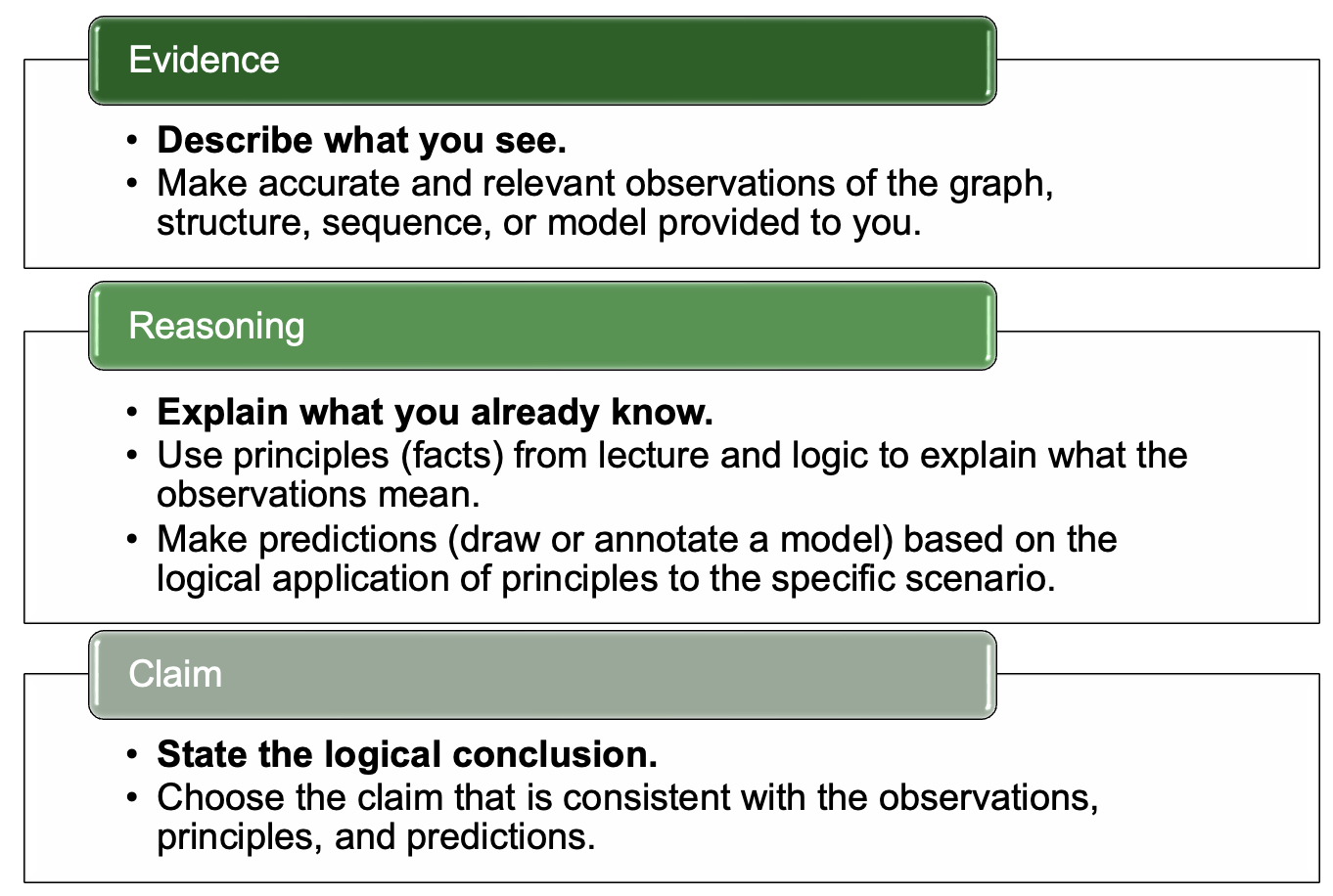
Evidence
The evidence is the results of an experiment. For our purposes, in addition to graphs or other types of data from an experiment, you may be presented with chemical structures, sequences, or models. The observations of this evidence are accurate and relevant descriptions of this evidence. For example, if you were provided with a graph, your evidence statement might compare the results for two conditions in that graph.
Reasoning
The reasoning includes scientific principles, or known facts, along with logic to explain how these facts relate to the observations and lead to the claim. Principles are usually ideas from textbooks or lectures and give context for the observations. For example, if the experiment is about cell division and the evidence gives the number of cells, a relevant principle would be about the process of cell division and how this affects the number of cells. Using logic, you could make predictions about proteins involved in cell division to explain the data obtained. All of these are reasoning to help make sense of the evidence and lead to the claim.
Claim
A claim is a conclusion about biological phenomenon that is based on the observations of evidence and reasoning. Importantly, it is not a guess, but rather an informed choice as to the most likely explanation that is consistent with known information. It helps to save writing the claim for last, after thinking through what you see from the evidence and what you know. As you are building your own arguments, be mindful that you are going where the evidence and reasoning leads you!
Linking Statement
To help reinforce the logical progression from observations to principles to the claim, our written arguments will include a linking statement. You can think of the linking statement as a “mini-argument” that includes all the elements of the scientific argument in about one sentence at the end of the argument. The linking statement will repeat (summarize) the key observations of the evidence and the key principles, and logically show how these lead to a specific claim.
Section 1.4 COVID Case Study
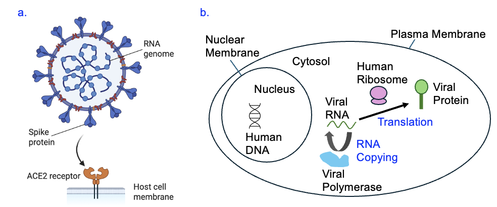
To begin our exploration of cells and molecules, we will focus on a topic that dominated our lives in recent years: COVID-19. COVID-19 is caused by the SARS-CoV-2 virus. Importantly, by the features of life that we have defined, virus meet some but not all the requirements, and thus are not considered living organisms. As shown in Figure 1.5a, SARS-CoV-2 does have a genome (although its sequence is RNA, not DNA), and is organized with biological molecules similarly to cells. However, a virus does not have cells or the ability to reproduce itself. Instead, it relies on host to do the work. In the case of SARS-CoV-2, the spike proteins that protrude from the surface of virus particle have the specific shape to interact with the ACE2 receptor, which is a protein found on host cells (Figure 1.5a). After binding to the host cell using this interaction, the virus inserts its RNA genome into the host cell and takes over gene expression in that cell. The host cell’s ribosomes use the viral RNA to produce viral proteins (Figure 1.5b), including all components of the virus. One of the viral proteins is a viral polymerase that copies the virus’s RNA genome. The viral components are assembled into virus particles that are then released from the host cell, ready to infect another host.
This is just a brief introduction to the virus to get you started on your pre-class preparation. We hope that you can bring your own knowledge and experiences to the discussion as well as learn from the knowledge of your peers, and use that as the foundation for biological inquiry!
